
Page 200 - Clinical Manual of Small Animal Endosurgery
P. 200
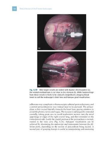
188   Clinical Manual of Small Animal Endosurgery


                                      (a)


                                      (b)

                              Fig. 6.10  After larger vessels are sealed with bipolar electrocautery (a),
                              the ventral mediastinum is cut close to the sternum (b). While haemorrhage
                              from these vessels is likely to be clinically insignificant, dripping blood
                              tends to soil the endoscope’s distal lens and hamper good visualisation.


                              adhesions may complicate a thoracoscopic subtotal pericardiectomy, and
                              a partial pericardiectomy may instead need to be pursued. The pericar-
                              dium is then incised laterally towards the heart base, paying attention to
                              avoid the phrenic nerves and vagus. The incision is subsequently extended
                              cranially, taking great care to avoid inadvertent incision into the atrial
                              appendage or edges of the right cranial lung, and then extended on the
                              contralateral side. Lastly the caudal portion of the pericardium is incised,
                              ventral  to  the  vena  cava  (Fig.  6.16).  Adequate  visualisation  can  be
                              achieved by alternating the endoscope and instruments between the dif-
                              ferent ports, depending on the section of pericardium being incised. A
                              second pair of grasping forceps is useful in manipulating and tensioning