
Page 204 - Clinical Manual of Small Animal Endosurgery
P. 204
192 Clinical Manual of Small Animal Endosurgery
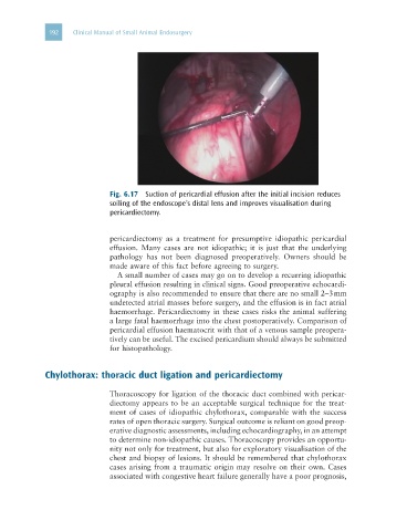
Fig. 6.17 Suction of pericardial effusion after the initial incision reduces
soiling of the endoscope’s distal lens and improves visualisation during
pericardiectomy.
pericardiectomy as a treatment for presumptive idiopathic pericardial
effusion. Many cases are not idiopathic; it is just that the underlying
pathology has not been diagnosed preoperatively. Owners should be
made aware of this fact before agreeing to surgery.
A small number of cases may go on to develop a recurring idiopathic
pleural effusion resulting in clinical signs. Good preoperative echocardi-
ography is also recommended to ensure that there are no small 2–3 mm
undetected atrial masses before surgery, and the effusion is in fact atrial
haemorrhage. Pericardiectomy in these cases risks the animal suffering
a large fatal haemorrhage into the chest postoperatively. Comparison of
pericardial effusion haematocrit with that of a venous sample preopera-
tively can be useful. The excised pericardium should always be submitted
for histopathology.
Chylothorax: thoracic duct ligation and pericardiectomy
Thoracoscopy for ligation of the thoracic duct combined with pericar-
diectomy appears to be an acceptable surgical technique for the treat-
ment of cases of idiopathic chylothorax, comparable with the success
rates of open thoracic surgery. Surgical outcome is reliant on good preop-
erative diagnostic assessments, including echocardiography, in an attempt
to determine non-idiopathic causes. Thoracoscopy provides an opportu-
nity not only for treatment, but also for exploratory visualisation of the
chest and biopsy of lesions. It should be remembered that chylothorax
cases arising from a traumatic origin may resolve on their own. Cases
associated with congestive heart failure generally have a poor prognosis,