
Page 207 - Clinical Manual of Small Animal Endosurgery
P. 207
Thoracoscopy 195
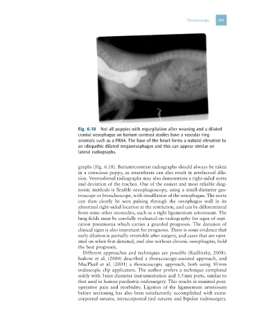
Fig. 6.18 Not all puppies with regurgitation after weaning and a dilated
cranial oesophagus on barium contrast studies have a vascular ring
anomaly such as a PRAA. The base of the heart forms a natural elevation to
an idiopathic dilated megaoesophagus and this can appear similar on
lateral radiographs.
graphs (Fig. 6.18). Barium/contrast radiographs should always be taken
in a conscious puppy, as anaesthesia can also result in artefactual dila-
tion. Ventrodorsal radiographs may also demonstrate a right-sided aorta
and deviation of the trachea. One of the easiest and most reliable diag-
nostic methods is flexible oesophagoscopy, using a small-diameter gas-
troscope or bronchoscope, with insufflation of the oesophagus. The aorta
can then clearly be seen pulsing through the oesophagus wall in its
abnormal right-sided location at the restriction, and can be differentiated
from some other anomalies, such as a right ligamentum arteriosum. The
lung fields must be carefully evaluated on radiography for signs of aspi-
ration pneumonia which carries a guarded prognosis. The duration of
clinical signs is also important for prognosis. There is some evidence that
early dilation is partially reversible after surgery, and cases that are oper-
ated on when first detected, and also without chronic oesophagitis, hold
the best prognosis.
Different approaches and techniques are possible (Radlinsky, 2008).
Isakow et al. (2000) described a thoracoscopy-assisted approach, and
MacPhail et al. (2001) a thoracoscopic approach, both using 10 mm
endoscopic clip applicators. The author prefers a technique completed
solely with 3 mm diameter instrumentation and 3.5 mm ports, similar to
that used in human paediatric endosurgery. This results in minimal post-
operative pain and morbidity. Ligation of the ligamentum arteriosum
before sectioning has also been satisfactorily accomplished with extra-
corporeal sutures, intracorporeal tied sutures and bipolar radiosurgery.