
Page 212 - Clinical Manual of Small Animal Endosurgery
P. 212
200 Clinical Manual of Small Animal Endosurgery
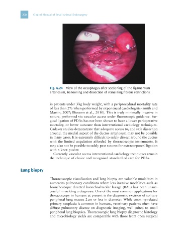
Fig. 6.24 View of the oesophagus after sectioning of the ligamentum
arteriosum, ballooning and dissection of remaining fibrous restrictions.
in patients under 3 kg body weight, with a periprocedural mortality rate
of less than 2% when performed by experienced cardiologists (Smith and
Martin, 2007; Blossom et al., 2010). This is truly minimally invasive in
nature, performed via vascular access under fluoroscopic guidance. Sur-
gical ligation of PDAs has not been shown to have a lower perioperative
mortality, or better outcome than interventional cardiology techniques.
Cadaver studies demonstrate that adequate access to, and safe dissection
around, the medial aspect of the ductus arteriosum may not be possible
in many cases. It is extremely difficult to safely dissect around the ductus
with the limited angulation afforded by thoracoscopic instruments. It
may also not be possible to safely pass sutures for extracorporeal ligation
with a knot pusher.
Currently vascular access interventional cardiology techniques remain
the technique of choice and recognised standard of care for PDAs.
Lung biopsy
Thoracoscopic visualisation and lung biopsy are valuable modalities in
numerous pulmonary conditions where less invasive modalities such as
bronchoscopic directed bronchoalveolar lavage (BAL) has been unsuc-
cessful in yielding a diagnosis. One of the most common applications for
thoracoscopy in humans at present is the diagnostic excision of solitary
peripheral lung masses 2 cm or less in diameter. While smoking-related
primary neoplasia is common in humans, veterinary patients often have
diffuse pulmonary disease on diagnostic imaging, well suited to small
peripheral lung biopsies. Thoracoscopic lung biopsy diagnostic histology
and microbiology yields are comparable with those from open surgical