
Page 208 - Clinical Manual of Small Animal Endosurgery
P. 208
196 Clinical Manual of Small Animal Endosurgery
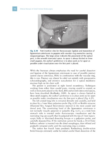
Fig. 6.19 Port-insertion sites for thoracoscopic ligation and transection of
ligamentum arteriosum in puppies with vascular ring anomalies causing
megaoesophagus. The large arrow indicates the placement of the optical
port, in the seventh intercostal space. As space is always limited in these
small puppies, the author’s preference is to place ports as far apart as
possible under visualisation once the first port is placed.
While the literature always emphasises the need for careful dissection
and ligation of the ligamentum arteriosum in case of possible patency
(patent ductus arteriosus, PDA) in combination with the vascular ring,
this is rare. Patency can almost be ruled out entirely with preoperative
echocardiography, and attentive auscultation for a typical machinery
murmur high on the chest wall.
The patient is positioned in right lateral recumbency. Techniques
working from either three caudal ports, viewing caudal to cranial, as
well as from ports placed at the third, fifth and seventh intercostal spaces,
have been described (Radlinsky, 2008). As space is always limited in
these small puppies, the author’s preference is to place ports as far apart
as possible under visualisation once the first port is placed (Fig. 6.19).
The left cranial lung lobe is retracted dorsally and caudally, and held
in place by a 3 mm blunt palpation probe (Fig. 6.20) or flexible retractor
(Diamond flex, Surgical Innovations) inserted in the most caudal and
dorsal port. The constricting band of the ligamentum arteriosum is
not normally visually apparent or palpable (Fig. 6.21), so a wide-bore
stomach tube is inserted under thoracoscopic visualisation and the
restricting ring can usually then be palpated with the tips of 3 mm laparo-
scopic Kelly or Maryland dissecting forceps or a palpation probe, and
carefully dissected free. If the restriction cannot be found, a 30 ml Foley
balloon catheter is inserted into the distal oesophagus, then inflated and
gradually retracted orally until the restricting ring is evident.
The author has found 3 mm paediatric Rothenburg double-action
bowel forceps extremely useful for initial careful blunt dissection of the