
Page 209 - Clinical Manual of Small Animal Endosurgery
P. 209
Thoracoscopy 197
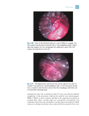
Fig. 6.20 View of the left heart base in a case of PRAA in a puppy. The
left cranial lung has been retracted with a 3 mm palpation probe. Labels
show the azygous vein (A), oesophagus (O), pulmonary artery (PA), left
atrium (LA) and phrenic nerve (P).
Fig. 6.21 The ligamentum arteriosum may not be apparent in cases of
vascular ring anomalies. Careful palpation with a 3 mm dissection forceps
once a stomach tube has been placed into the oesophagus will allow one
to detect the restricting band.
mediastinum after this is initially incised. In some cases this has allowed
completion of the procedure without the need for any electrosurgery.
While a PRAA is most common and this is the anatomical position to
examine first, other vascular rings are occasionally encountered, and
sometimes more than one anomalous vascular ring is encountered. Small
tortuous overlying vessels may also commonly be encountered. Even very