
Page 210 - Clinical Manual of Small Animal Endosurgery
P. 210
198 Clinical Manual of Small Animal Endosurgery
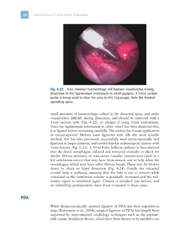
Fig. 6.22 Even minimal haemorrhage will hamper visualisation during
dissection of the ligamentum arteriosum in small puppies. A 3 mm suction
probe is being used to clear the area in this 3 kg puppy. Note the limited
operating space.
small amounts of haemorrhage collect in the dissected space and make
visualisation difficult during dissection, and should be removed with a
3 mm suction tube (Fig. 6.22), or pledget if using 5 mm instruments.
Once the ligamentum arteriosum or other vessel has been dissected free,
it is ligated before sectioning carefully. The author has found application
of extracorporeal Meltzer knot ligatures with silk the most reliable
method, but has also previously successfully used intracorporeally tied
ligatures in larger patients, and careful bipolar radiosurgical cautery with
3 mm forceps (Fig. 6.23). A 30 ml Foley balloon catheter is then inserted
into the distal oesophagus, inflated and retracted cranially to check for
further fibrous strictures or concurrent vascular constrictions (such as a
left subclavian artery) that may have been missed, and to help dilate the
oesophagus, which may have other fibrous bands. These may be broken
down by sharp or blunt dissection (Fig. 6.24). Finally the retracted
cranial lung is replaced, ensuring that the lobe is not in torsion while
visualised as the ventilation volume is gradually increased and the atel-
ectatic region is ventilated again. Closure is standard (see below), and
no indwelling postoperative chest drain is needed in these cases.
PDA
While thoracoscopically assisted ligation of PDA has been reported in
dogs (Borenstein et al., 2004), surgical ligation of PDAs has largely been
surpassed by interventional cardiology techniques such as the implant-
able canine Amplatzer device, which have been shown to be suitable even