
Page 241 - Clinical Manual of Small Animal Endosurgery
P. 241
Female Reproductive Tract 229
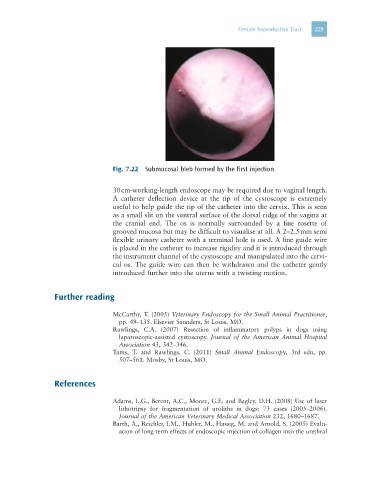
Fig. 7.22 Submucosal bleb formed by the first injection.
30 cm-working-length endoscope may be required due to vaginal length.
A catheter deflection device at the tip of the cystoscope is extremely
useful to help guide the tip of the catheter into the cervix. This is seen
as a small slit on the ventral surface of the dorsal ridge of the vagina at
the cranial end. The os is normally surrounded by a fine rosette of
grooved mucosa but may be difficult to visualise at all. A 2–2.5 mm semi
flexible urinary catheter with a terminal hole is used. A fine guide wire
is placed in the catheter to increase rigidity and it is introduced through
the instrument channel of the cystoscope and manipulated into the cervi-
cal os. The guide wire can then be withdrawn and the catheter gently
introduced further into the uterus with a twisting motion.
Further reading
McCarthy, T. (2005) Veterinary Endoscopy for the Small Animal Practitioner,
pp. 49–135. Elsevier Saunders, St Louis, MO.
Rawlings, C.A. (2007) Resection of inflammatory polyps in dogs using
laparoscopic-assisted cystoscopy. Journal of the American Animal Hospital
Association 43, 342–346.
Tams, T. and Rawlings, C. (2011) Small Animal Endoscopy, 3rd edn, pp.
507–561. Mosby, St Louis, MO.
References
Adams, L.G., Berent, A.C., Moore, G.E. and Bagley, D.H. (2008) Use of laser
lithotripsy for fragmentation of uroliths in dogs: 73 cases (2005–2006).
Journal of the American Veterinary Medical Association 232, 1680–1687.
Barth, A., Reichler, I.M., Hubler, M., Hassig, M. and Arnold, S. (2005) Evalu-
ation of long-term effects of endoscopic injection of collagen into the urethral