
Page 246 - Clinical Manual of Small Animal Endosurgery
P. 246
234 Clinical Manual of Small Animal Endosurgery
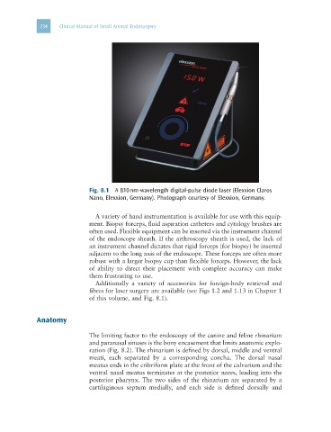
Fig. 8.1 A 810 nm-wavelength digital-pulse diode laser (Elexxion Claros
Nano, Elexxion, Germany). Photograph courtesy of Elexxion, Germany.
A variety of hand instrumentation is available for use with this equip-
ment. Biopsy forceps, fluid aspiration catheters and cytology brushes are
often used. Flexible equipment can be inserted via the instrument channel
of the endoscope sheath. If the arthroscopy sheath is used, the lack of
an instrument channel dictates that rigid forceps (for biopsy) be inserted
adjacent to the long axis of the endoscope. These forceps are often more
robust with a larger biopsy cup than flexible forceps. However, the lack
of ability to direct their placement with complete accuracy can make
them frustrating to use.
Additionally a variety of accessories for foreign-body retrieval and
fibres for laser surgery are available (see Figs 1.2 and 1.13 in Chapter 1
of this volume, and Fig. 8.1).
Anatomy
The limiting factor to the endoscopy of the canine and feline rhinarium
and paranasal sinuses is the bony encasement that limits anatomic explo-
ration (Fig. 8.2). The rhinarium is defined by dorsal, middle and ventral
meati, each separated by a corresponding concha. The dorsal nasal
meatus ends in the cribriform plate at the front of the calvarium and the
ventral nasal meatus terminates at the posterior nares, leading into the
posterior pharynx. The two sides of the rhinarium are separated by a
cartilaginous septum medially, and each side is defined dorsally and