
Page 248 - Clinical Manual of Small Animal Endosurgery
P. 248
236 Clinical Manual of Small Animal Endosurgery
laterally by the nasal and frontal bones, laterally and ventrally by the
maxillary and palatine bones and rostrally by the nasal planum. The
nostrils serve as the aperture into the nasal vestibule. The puncta of the
nasolacrimal duct are located in the nasal vestibule along the most
ventral aspect of the alar cartilage.
The paranasal sinuses are a series of bilateral, somewhat intercon-
nected, air-filled spaces lined with a highly secretory mucous membrane.
As a practical matter, the most clinically significant of the paranasal
sinuses is the frontal sinus, which can be accessed endoscopically.
The mucous membrane of the rhinarium extending to the proximal
pharynx comprises a ciliated columnar epithelium. These highly vascular
membranes, draped over the tremendous surface area of the nasal
conchae, serve to warm, humidify and filter the inspired air.
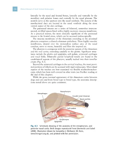
The pharynx is contiguous with the posterior aspects of the rhinarium
and the oral cavity, extending caudally to the glottis. Significant struc-
tures include the glottis and epiglottis, soft palate, arytenoid cartilages
and vocal folds. Bilaterally paired lymphoid tonsils are found in the
caudolateral aspects of the pharynx, usually tucked into their tonsillar
crypts (Fig. 8.3).
Beyond the arytenoid cartilages is the cervical trachea, the most proxi-
mal portions of which can be accessed with rigid endoscopes. More distal
aspects of the trachea are best examined via flexible tracheobronchos-
copy, which has been well covered in other texts (see Further reading at
the end of this chapter).
While the gross external appearance of the rhinarium varies between
dogs and cats and from breed type to breed type, the anatomic descrip-
tions noted above are quite consistent.
Nasopharynx
Oesophagus
Caudal nasal choanae
Nasal cavities
Soft palate
Tongue
Trachea
Oropharynx
Epiglottis
Fig. 8.3 Schematic drawing of the anatomy of the retropharynx. and
posterior nasal cavity. Both images reproduced from Lhermette and Sobel
(2008). Illustration drawn by Samantha J. Elmhurst, BA Hons,
www.livingart.org.uk, and printed with her permission.