
Page 252 - Clinical Manual of Small Animal Endosurgery
P. 252
240 Clinical Manual of Small Animal Endosurgery
(a) (b) (c)
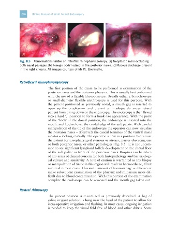
Fig. 8.5 Abnormalities visible on retroflex rhinopharyngoscopy. (a) Neoplastic mass occluding
both nasal passages. (b) Foreign body lodged in the posterior nares. (c) Mucous discharge present
in the right choana. All images courtesy of Mr P.J. Lhermette.
Retroflexed rhinopharyngoscopy
The first portion of the exam to be performed is examination of the
posterior nares and the posterior pharynx. This is usually best performed
with the use of a flexible fibreoptiscope. Usually either a bronchoscope
or small-diameter flexible urethroscope is used for this purpose. With
the patient positioned as previously noted, a mouth gag is inserted to
open up the oropharynx and prevent an inadequately anaesthetised
patient from biting down on the endoscope. The endoscope is then flexed
into a hard ‘J’ position to form a hook-like appearance. With the point
of the ‘hook’ in the dorsal position, the endoscope is inserted into the
mouth and hooked over the caudal edge of the soft palate. With careful
manipulation of the tip of the endoscope the operator can now visualise
the posterior nares – effectively the caudal terminus of the ventral nasal
meatus – looking rostrally. The operator is now in a position to examine
the patient for nasopharyngeal stenosis or atresia, masses obscuring one
or both posterior nares, or other pathologies (Fig. 8.5). It is not uncom-
mon to see significant lymphoid follicle development on the dorsal floor
of the soft palate in front of the posterior nares. Biopsies can be taken
of any areas of clinical concern for both histopathology and bacteriologi-
cal culture and sensitivity. A note of caution is warranted as any biopsy
or manipulation of tissue in this region will result in haemorrhage, albeit
minimal in most cases. This small amount of haemorrhage will however
make subsequent examination of the pharynx and rhinarium more dif-
ficult due to blood contamination. With this portion of the examination
complete the endoscope can be removed and the mouth gag taken out.
Rostral rhinoscopy
The patient position is maintained as previously described. A bag of
saline irrigant solution is hung near the head of the patient to allow for
intra-operative irrigation and flushing. In most cases, ongoing irrigation
is needed to keep the visual field free of blood and other debris. Some