
Page 253 - Clinical Manual of Small Animal Endosurgery
P. 253
Upper Respiratory Tract 241
authors have advocated using cool saline and others have suggested the
use of dilute adrenaline (epinephrine) or other vasoconstrictive agents in
an effort to minimise haemorrhage. This author has not found it neces-
sary to do so.
As previously noted, this author performs the vast majority of rhinos-
copy with the 2.7 mm, 30° urethrocystoscope. However, there are some
limited circumstances where the small-diameter flexible scope is of value.
Small patients or lesions that require odd angulation to visualise ade-
quately may benefit from the two-way deflection afforded by the flexible
endoscope.
The rigid endoscope is inserted into the nose via the nostril across the
alar cartilage. Generally speaking a slight dorsal deflection of the tip of
the endoscope is needed to get over the ventral ridge of the alar cartilage.
At this point, fluid irrigation should be begun. It bears noting that even
minimal, seemingly innocuous manipulation of the nasal mucosa will
cause some haemorrhage. While usually of no clinical significance, the
blood can make keeping the field of view clear more difficult. Care
should consequently be taken with all intraluminal manipulations.
A systematic examination of the rhinarium should be undertaken.
This author usually starts with examination of the dorsal nasal meatus
and concha. While it is usually difficult to examine the dorsal meatus to
the level of the cribriform plate, significant posterior progress should be
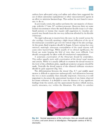
made. Ventrally the dorsal aspects of the ethmoid turbinate structures
should be quite apparent (Fig. 8.6).
The differentiation between the dorsal (Fig. 8.7) and middle nasal
meatus is difficult to appreciate endoscopically and delineation between
the two is more academic than clinically important. However, it is still
important to document the approximate location of any lesion observed
for future reference. It is helpful to note the distance of any significant
findings from the nostril, and the approximate location (which meatus,
nearby structures, etc.) within the rhinarium. The ability to correlate
Fig. 8.6 Normal appearance of the turbinates: they are smooth and pink
in colour, and seem almost to interdigitate. Photograph courtesy of Mr P.J.
Lhermette.