
Page 258 - Clinical Manual of Small Animal Endosurgery
P. 258
246 Clinical Manual of Small Animal Endosurgery
(a) (b) (c)
Fig. 8.9 (a) Benign polyp in the choanae as it appears with retroflex rhinopharyngoscopy. (b)
Polyp seen at anterior rhinoscopy. In this case there is a solitary polyp, confined to a small area,
whereas in (c) the polyp occupies most of the nasal passage. All images reproduced from
Lhermette and Sobel (2008), with the permission of BSAVA publications. © BSAVA.
(a) (b) (c)
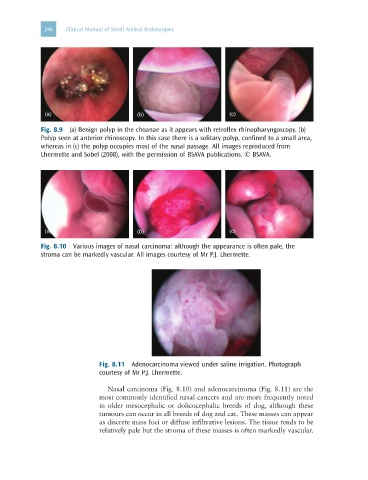
Fig. 8.10 Various images of nasal carcinoma: although the appearance is often pale, the
stroma can be markedly vascular. All images courtesy of Mr P.J. Lhermette.
Fig. 8.11 Adenocarcinoma viewed under saline irrigation. Photograph
courtesy of Mr P.J. Lhermette.
Nasal carcinoma (Fig. 8.10) and adenocarcinoma (Fig. 8.11) are the
most commonly identified nasal cancers and are more frequently noted
in older mesocephalic or dolicocephalic breeds of dog, although these
tumours can occur in all breeds of dog and cat. These masses can appear
as discrete mass foci or diffuse infiltrative lesions. The tissue tends to be
relatively pale but the stroma of these masses is often markedly vascular.