
Page 259 - Clinical Manual of Small Animal Endosurgery
P. 259
Upper Respiratory Tract 247
It is worth noting that it is often difficult to identify all the margins and
anatomic limitations of these lesions at rhinoscopy. As previously dis-
cussed, a combination of radiography and/or CT or MRI will help the
surgeon delineate the extent of the mass. These tumours are known to
be markedly sensitive to radiation therapy and both palliative and cura-
tive protocols are described. The diode laser has also been shown to have
significant palliative benefit, but should not be regarded as curative or
as having a long-term management effect. In patients for whom radiation
therapy is not an option, this author often uses a combination of laser
ablation and oral piroxicam as a palliative management strategy.
Fungal disease
The most common fungal disease observed in the rhinarium and para-
nasal sinuses is aspergillosis. Aspergillus is a soil-borne environmental
pathogen that can cause chronic destructive mycotic rhinitis in the nose
and sinuses of, most commonly, sporting dogs, although any breed or
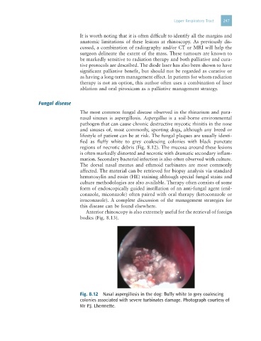
lifestyle of patient can be at risk. The fungal plaques are usually identi-
fied as fluffy white to grey coalescing colonies with black punctate
regions of necrotic debris (Fig. 8.12). The mucosa around these lesions
is often markedly distorted and necrotic with dramatic secondary inflam-
mation. Secondary bacterial infection is also often observed with culture.
The dorsal nasal meatus and ethmoid turbinates are most commonly
affected. The material can be retrieved for biopsy analysis via standard
hematoxylin and eosin (HE) staining although special fungal stains and
culture methodologies are also available. Therapy often consists of some
form of endoscopically guided instillation of an anti-fungal agent (enil-
conazole, miconazole) often paired with oral therapy (ketoconazole or
itraconazole). A complete discussion of the management strategies for
this disease can be found elsewhere.
Anterior rhinoscopy is also extremely useful for the retrieval of foreign
bodies (Fig. 8.13).
Fig. 8.12 Nasal aspergillosis in the dog: fluffy white to grey coalescing
colonies associated with severe turbinates damage. Photograph courtesy of
Mr P.J. Lhermette.