
Page 264 - Clinical Manual of Small Animal Endosurgery
P. 264
252 Clinical Manual of Small Animal Endosurgery
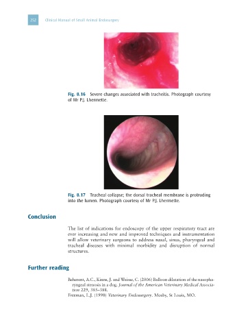
Fig. 8.16 Severe changes associated with tracheitis. Photograph courtesy
of Mr P.J. Lhermette.
Fig. 8.17 Tracheal collapse; the dorsal tracheal membrane is protruding
into the lumen. Photograph courtesy of Mr P.J. Lhermette.
Conclusion
The list of indications for endoscopy of the upper respiratory tract are
ever increasing and new and improved techniques and instrumentation
will allow veterinary surgeons to address nasal, sinus, pharyngeal and
tracheal diseases with minimal morbidity and disruption of normal
structures.
Further reading
Beherent, A.C., Kinns, J. and Weisse, C. (2006) Balloon dilatation of the nasopha-
ryngeal stenosis in a dog. Journal of the American Veterinary Medical Associa-
tion 229, 385–388.
Freeman, L.J. (1998) Veterinary Endosurgery. Mosby, St Louis, MO.