
Page 269 - Clinical Manual of Small Animal Endosurgery
P. 269
Otoendoscopy 257
While this author does use 0° otoendoscopes for some diagnostic
otoendoscopy, the use of a 2.7 mm, 30° rigid endoscope is preferable for
working on larger canine patients and doing most interventional proce-
dures. These endoscopes are often marketed as ‘multipurpose rigid endo-
scopes’ but are essentially paediatric urethrocystoscopes in an appropriate
sheath (see Fig. 1.2). The advantages to these endoscopes are the increased
working length which allows for complete visualisation of the horizontal
ear canal in even the largest patients, as well as the increased field of
view due to the 30° optical view. This is particularly advantageous in
visualising all areas of the ear canal at the level of the junction of its
vertical and horizontal portions as well as providing complete visualisa-
tion of the tympanum. In addition, these endoscopes have separate
ingress and egress channels allowing for adequate fluid irrigation and
drainage as well as a separate instrument channel.
A full range of cytology, culture and cleaning brushes as well as
curettes, biopsy forceps and graspers are available. The author also uses
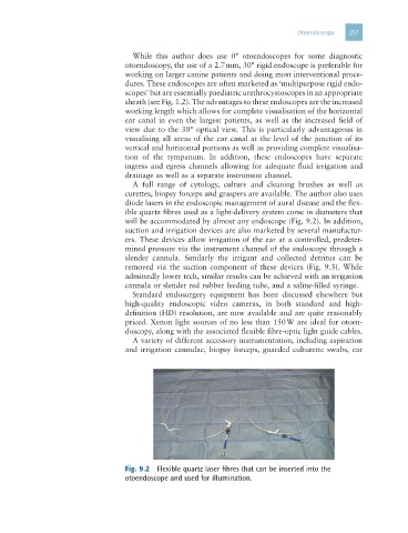
diode lasers in the endoscopic management of aural disease and the flex-
ible quartz fibres used as a light-delivery system come in diameters that
will be accommodated by almost any endoscope (Fig. 9.2). In addition,
suction and irrigation devices are also marketed by several manufactur-
ers. These devices allow irrigation of the ear at a controlled, predeter-
mined pressure via the instrument channel of the endoscope through a
slender cannula. Similarly the irrigant and collected detritus can be
removed via the suction component of these devices (Fig. 9.3). While
admittedly lower tech, similar results can be achieved with an irrigation
cannula or slender red rubber feeding tube, and a saline-filled syringe.
Standard endosurgery equipment has been discussed elsewhere but
high-quality endoscopic video cameras, in both standard and high-
definition (HD) resolution, are now available and are quite reasonably
priced. Xenon light sources of no less than 150 W are ideal for otoen-
doscopy, along with the associated flexible fibre-optic light guide cables.
A variety of different accessory instrumentation, including aspiration
and irrigation cannulae, biopsy forceps, guarded culturette swabs, ear
Fig. 9.2 Flexible quartz laser fibres that can be inserted into the
otoendoscope and used for illumination.