
Page 273 - Clinical Manual of Small Animal Endosurgery
P. 273
Otoendoscopy 261
(a)
(b)
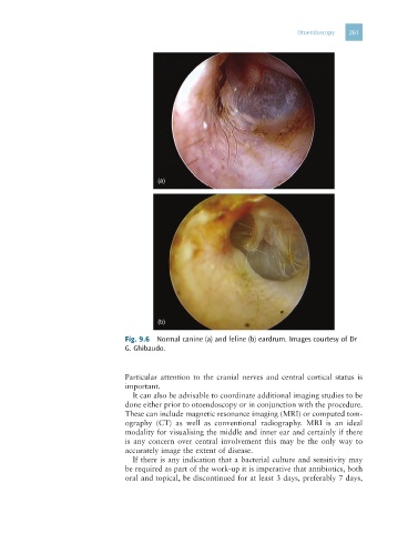
Fig. 9.6 Normal canine (a) and feline (b) eardrum. Images courtesy of Dr
G. Ghibaudo.
Particular attention to the cranial nerves and central cortical status is
important.
It can also be advisable to coordinate additional imaging studies to be
done either prior to otoendoscopy or in conjunction with the procedure.
These can include magnetic resonance imaging (MRI) or computed tom-
ography (CT) as well as conventional radiography. MRI is an ideal
modality for visualising the middle and inner ear and certainly if there
is any concern over central involvement this may be the only way to
accurately image the extent of disease.
If there is any indication that a bacterial culture and sensitivity may
be required as part of the work-up it is imperative that antibiotics, both
oral and topical, be discontinued for at least 3 days, preferably 7 days,