
Page 274 - Clinical Manual of Small Animal Endosurgery
P. 274
262 Clinical Manual of Small Animal Endosurgery
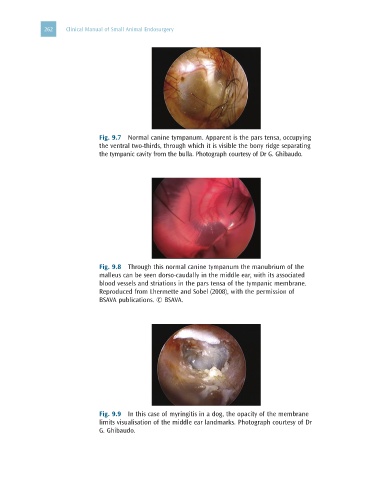
Fig. 9.7 Normal canine tympanum. Apparent is the pars tensa, occupying
the ventral two-thirds, through which it is visible the bony ridge separating
the tympanic cavity from the bulla. Photograph courtesy of Dr G. Ghibaudo.
Fig. 9.8 Through this normal canine tympanum the manubrium of the
malleus can be seen dorso-caudally in the middle ear, with its associated
blood vessels and striations in the pars tensa of the tympanic membrane.
Reproduced from Lhermette and Sobel (2008), with the permission of
BSAVA publications. © BSAVA.
Fig. 9.9 In this case of myringitis in a dog, the opacity of the membrane
limits visualisation of the middle ear landmarks. Photograph courtesy of Dr
G. Ghibaudo.