
Page 279 - Clinical Manual of Small Animal Endosurgery
P. 279
Otoendoscopy 267
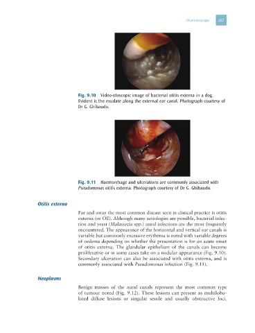
Fig. 9.10 Video-otoscopic image of bacterial otitis externa in a dog.
Evident is the exudate along the external ear canal. Photograph courtesy of
Dr G. Ghibaudo.
Fig. 9.11 Haemorrhage and ulcerations are commonly associated with
Pseudomonas otitis externa. Photograph courtesy of Dr G. Ghibaudo.
Otitis externa
Far and away the most common disease seen in clinical practice is otitis
externa (or OE). Although many aetiologies are possible, bacterial infec-
tion and yeast (Malassezia spp.) aural infections are the most frequently
encountered. The appearance of the horizontal and vertical ear canals is
variable but commonly excessive erythema is noted with variable degrees
of oedema depending on whether the presentation is for an acute onset
of otitis externa. The glandular epithelium of the canals can become
proliferative or in some cases take on a nodular appearance (Fig. 9.10).
Secondary ulceration can also be associated with otitis externa, and is
commonly associated with Pseudomonas infection (Fig. 9.11).
Neoplasms
Benign masses of the aural canals represent the most common type
of tumour noted (Fig. 9.12). These lesions can present as multilobu-
lated diffuse lesions or singular sessile and usually obstructive foci.