
Page 281 - Clinical Manual of Small Animal Endosurgery
P. 281
Otoendoscopy 269
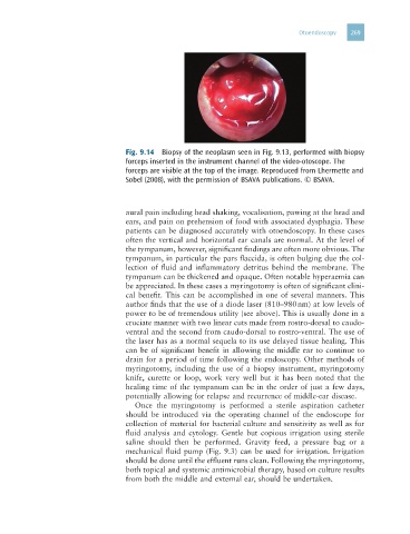
Fig. 9.14 Biopsy of the neoplasm seen in Fig. 9.13, performed with biopsy
forceps inserted in the instrument channel of the video-otoscope. The
forceps are visible at the top of the image. Reproduced from Lhermette and
Sobel (2008), with the permission of BSAVA publications. © BSAVA.
aural pain including head shaking, vocalisation, pawing at the head and
ears, and pain on prehension of food with associated dysphagia. These
patients can be diagnosed accurately with otoendoscopy. In these cases
often the vertical and horizontal ear canals are normal. At the level of
the tympanum, however, significant findings are often more obvious. The
tympanum, in particular the pars flaccida, is often bulging due the col-
lection of fluid and inflammatory detritus behind the membrane. The
tympanum can be thickened and opaque. Often notable hyperaemia can
be appreciated. In these cases a myringotomy is often of significant clini-
cal benefit. This can be accomplished in one of several manners. This
author finds that the use of a diode laser (810–980 nm) at low levels of
power to be of tremendous utility (see above). This is usually done in a
cruciate manner with two linear cuts made from rostro-dorsal to caudo-
ventral and the second from caudo-dorsal to rostro-ventral. The use of
the laser has as a normal sequela to its use delayed tissue healing. This
can be of significant benefit in allowing the middle ear to continue to
drain for a period of time following the endoscopy. Other methods of
myringotomy, including the use of a biopsy instrument, myringotomy
knife, curette or loop, work very well but it has been noted that the
healing time of the tympanum can be in the order of just a few days,
potentially allowing for relapse and recurrence of middle-ear disease.
Once the myringotomy is performed a sterile aspiration catheter
should be introduced via the operating channel of the endoscope for
collection of material for bacterial culture and sensitivity as well as for
fluid analysis and cytology. Gentle but copious irrigation using sterile
saline should then be performed. Gravity feed, a pressure bag or a
mechanical fluid pump (Fig. 9.3) can be used for irrigation. Irrigation
should be done until the effluent runs clean. Following the myringotomy,
both topical and systemic antimicrobial therapy, based on culture results
from both the middle and external ear, should be undertaken.