
Page 280 - Clinical Manual of Small Animal Endosurgery
P. 280
268 Clinical Manual of Small Animal Endosurgery
(a) (b) (c)
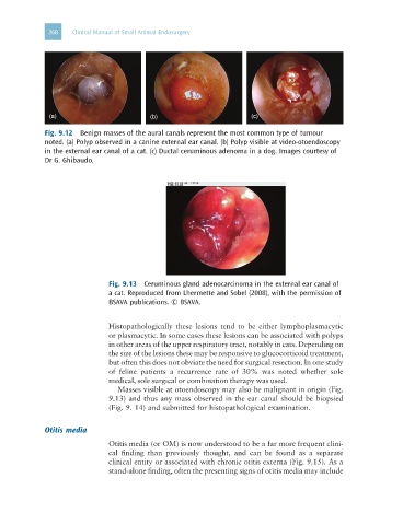
Fig. 9.12 Benign masses of the aural canals represent the most common type of tumour
noted. (a) Polyp observed in a canine external ear canal. (b) Polyp visible at video-otoendoscopy
in the external ear canal of a cat. (c) Ductal ceruminous adenoma in a dog. Images courtesy of
Dr G. Ghibaudo.
Fig. 9.13 Ceruminous gland adenocarcinoma in the external ear canal of
a cat. Reproduced from Lhermette and Sobel (2008), with the permission of
BSAVA publications. © BSAVA.
Histopathologically these lesions tend to be either lymphoplasmacytic
or plasmacytic. In some cases these lesions can be associated with polyps
in other areas of the upper respiratory tract, notably in cats. Depending on
the size of the lesions these may be responsive to glucocorticoid treatment,
but often this does not obviate the need for surgical resection. In one study
of feline patients a recurrence rate of 30% was noted whether sole
medical, sole surgical or combination therapy was used.
Masses visible at otoendoscopy may also be malignant in origin (Fig.
9.13) and thus any mass observed in the ear canal should be biopsied
(Fig. 9. 14) and submitted for histopathological examination.
Otitis media
Otitis media (or OM) is now understood to be a far more frequent clini-
cal finding than previously thought, and can be found as a separate
clinical entity or associated with chronic otitis externa (Fig. 9.15). As a
stand-alone finding, often the presenting signs of otitis media may include