
Page 283 - Clinical Manual of Small Animal Endosurgery
P. 283
Otoendoscopy 271
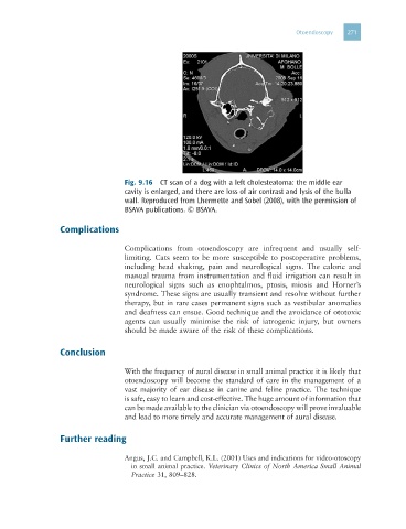
Fig. 9.16 CT scan of a dog with a left cholesteatoma: the middle ear
cavity is enlarged, and there are loss of air contrast and lysis of the bulla
wall. Reproduced from Lhermette and Sobel (2008), with the permission of
BSAVA publications. © BSAVA.
Complications
Complications from otoendoscopy are infrequent and usually self-
limiting. Cats seem to be more susceptible to postoperative problems,
including head shaking, pain and neurological signs. The caloric and
manual trauma from instrumentation and fluid irrigation can result in
neurological signs such as enophtalmos, ptosis, miosis and Horner’s
syndrome. These signs are usually transient and resolve without further
therapy, but in rare cases permanent signs such as vestibular anomalies
and deafness can ensue. Good technique and the avoidance of ototoxic
agents can usually minimise the risk of iatrogenic injury, but owners
should be made aware of the risk of these complications.
Conclusion
With the frequency of aural disease in small animal practice it is likely that
otoendoscopy will become the standard of care in the management of a
vast majority of ear disease in canine and feline practice. The technique
is safe, easy to learn and cost-effective. The huge amount of information that
can be made available to the clinician via otoendoscopy will prove invaluable
and lead to more timely and accurate management of aural disease.
Further reading
Angus, J.C. and Campbell, K.L. (2001) Uses and indications for video-otoscopy
in small animal practice. Veterinary Clinics of North America Small Animal
Practice 31, 809–828.