
Page 268 - Clinical Manual of Small Animal Endosurgery
P. 268
256 Clinical Manual of Small Animal Endosurgery
modality to therapeutically correct underlying pathology (tumour or
polyp resection with laser or electrocautery, foreign-body retrieval, deep-
ear cleaning and flushing, myringotomy). As such, otoendoscopy is a tech-
nique that is valuable for every endoscopist to have in their arsenal.
Indications
The clinical presentations of the patient with aural disease are all too
familiar to most veterinary practitioners. Ear shaking or aural pruritis,
chronic aural odour and/or discharge, aural pain, hearing loss and
peripheral neurological signs consistent with middle-ear disease are all
common presenting problems for the patient in need of otoendoscopy.
In addition, the clinical progress of patients with confirmed aural pathol-
ogy can be monitored. Patients with aural disease refractory to therapy
can also be evaluated and subsequent treatment plans modified to achieve
optimal clinical results.
Instrumentation
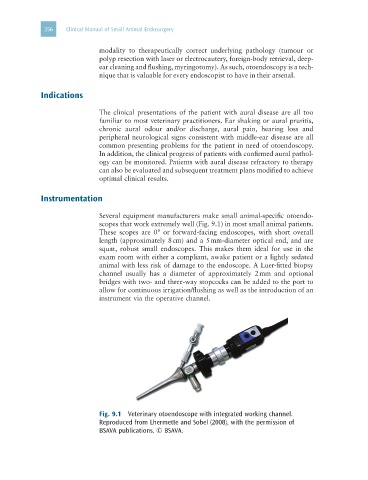
Several equipment manufacturers make small animal-specific otoendo-
scopes that work extremely well (Fig. 9.1) in most small animal patients.
These scopes are 0° or forward-facing endoscopes, with short overall
length (approximately 8 cm) and a 5 mm-diameter optical end, and are
squat, robust small endoscopes. This makes them ideal for use in the
exam room with either a compliant, awake patient or a lightly sedated
animal with less risk of damage to the endoscope. A Luer-fitted biopsy
channel usually has a diameter of approximately 2 mm and optional
bridges with two- and three-way stopcocks can be added to the port to
allow for continuous irrigation/flushing as well as the introduction of an
instrument via the operative channel.
Fig. 9.1 Veterinary otoendoscope with integrated working channel.
Reproduced from Lhermette and Sobel (2008), with the permission of
BSAVA publications. © BSAVA.