
Page 263 - Clinical Manual of Small Animal Endosurgery
P. 263
Upper Respiratory Tract 251
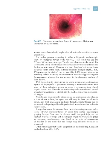
Fig. 8.15 Trachea as seen using a 5 mm, 0° laparoscope. Photograph
courtesy of Mr P.J. Lhermette.
intravenous catheter should be placed to allow for the use of intravenous
anaesthetics.
For smaller patients presenting for either a diagnostic tracheoscopic
exam or emergency foreign body removal, I can sometimes use the
2.7 mm, 30° urethrocystoscope. The obvious advantage to the use of this
scope is the ability to introduce operative accessory instrumentation via
the instrument channel. However, the short length of this scope limits
the distal extent of the exam. In those situations a longer-length, 5 mm,
0° laparoscope (or similar) can be used (Fig. 8.15). Again, without an
operating sheath, accessory instrumentation must be slipped alongside
the endoscope, allowing for less accuracy in the placement and use of
these devices.
With the patient in either sternal or lateral recumbency an induction
agent such as propofol is given intravenously. Care must be taken with
many of these induction agents, as apnea is a common-dose-related
sequela to their use. When the patient is adequately anaesthetised a nasal
or oral oxygen catheter is slipped into the trachea to provide supplemen-
tal oxygen.
Propofol can be continually administered via continuous-rate infusion
or intermittent boluses, but speed and efficiency in these procedures is
paramount. With endoscopic guidance, bronchoalveolar lavage can be
performed and cytological brushings obtained from the trachea and main
stem bronchi.
Foreign bodies can be retrieved from the trachea using standard endo-
scopic retrieval instrumentation or using long-shafted laparoscopic
grasping forceps. Care must be taken to avoid iatrogenic injury to the
tracheal mucosa or rings and the surgeon must be prepared to place
an emergency tracheostomy tube distal to the point of obstruction
(if possible) in the event that the foreign-body retrieval procedure is
prolonged.
Other pathologies that can be diagnosed are tracheitis (Fig. 8.16) and
tracheal collapse (Fig. 8.17).