
Page 261 - Clinical Manual of Small Animal Endosurgery
P. 261
Upper Respiratory Tract 249
use a slender solid quartz fibre as their delivery system. This makes them
ideal for introduction into the operating channel of the endoscope (be it
rigid or flexible). In addition, the wavelength of light that is produced
by most commercially available surgical diodes ranges from 810 to
980 nm. These wavelengths of light perform particularly well in fluid
mediums, such as those encountered in nasal endoscopic surgery. In
particular, diodes at the 810 nm wavelength are particularly well absorbed
by biological pigments (haemoglobin), thus maximising their thermal effect.
The diodes are used to vapourise and ablate abnormal tissue. This is
done by introducing a fibre of appropriate size into the area of interest via the
operating channel of the endoscope. A power level is then selected, a pulse
interval chosen and the fibre placed in apposition to the affected tissues. The
procedure is continued until all grossly identifiable abnormal tissue is removed
or until normal anatomical landmarks can no longer be easily identified.
The diode lasers have been used to manage many different types of
nasal pathology but their greatest utility is as adjunctive therapy for
neoplastic diseases. Often the procedure is performed as part of the initial
rhinoscopic examination in the hopes of reducing tumour volume for the
benefit of subsequent, more definitive therapy (e.g. radiation therapy).
However, we have performed many cases where laser surgery was the
sole therapy chosen by the owners, and good success has been noted. It
is clear that this procedure does not result in clean surgical margins, but
extended periods of control of clinical signs have been observed, often
in excess of 6 months. Lack of empirical data makes it difficult to recom-
mend laser surgery as a definitive therapy for nasal neoplasia, but its
benefit as an adjunctive or palliative modality is clear.
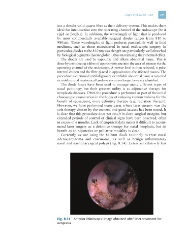
Currently we are using the 810 nm diode routinely to treat nasal
adenocarcinoma and carcinoma, as well as benign inflammatory
nasal and nasopharyngeal polyps (Fig. 8.14). Lasers are relatively less
Fig. 8.14 Anterior rhinoscopic image obtained after laser treatment for
neoplasia.