
Page 254 - Clinical Manual of Small Animal Endosurgery
P. 254
242 Clinical Manual of Small Animal Endosurgery
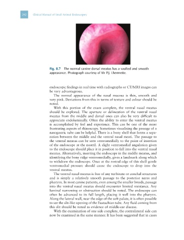
Fig. 8.7 The normal canine dorsal meatus has a vaulted and smooth
appearance. Photograph courtesy of Mr P.J. Lhermette.
endoscopic findings in real time with radiographs or CT/MRI images can
be very advantageous.
The normal appearance of the nasal mucosa is thin, smooth and
very pink. Deviations from this in terms of texture and colour should be
noted.
With this portion of the exam complete, the ventral nasal meatus
should be explored. The aperture or delineation of the ventral nasal
meatus from the middle and dorsal ones can also be very difficult to
appreciate endoluminally. Often the ability to enter the ventral meatus
is accomplished by feel and experience. This can be one of the more
frustrating aspects of rhinoscopy. Sometimes visualising the passage of a
nasogastric tube can be helpful. There is a bony shelf that forms a sepa-
ration between the middle and the ventral nasal meati. The passage to
the ventral meatus can be seen ventromedially to the point of insertion
of the endoscope at the nostril. A slight ventromedial angulation given
to the endoscope should place it in position to fall into the ventral nasal
meatus. Alternatively, inserting the endoscope in the middle meatus, and
identifying the bony ridge ventromedially, gives a landmark along which
to withdraw the endoscope. Once at the rostral edge of this shelf gentle
ventromedial pressure should cause the endoscope to drop into the
ventral meatus.
The ventral nasal meatus is free of any turbinate or conchal structures
and is simply a relatively smooth passage to the posterior nares and
pharynx. In most canine patients, even among the smaller breeds, passage
into the ventral nasal meatus should encounter limited resistance. Any
luminal narrowing or obstruction should be noted. The endoscope can
often be advanced to its full length, placing it well into the pharynx.
Along the lateral wall, near the edge of the soft palate, it is often possible
to see the slit-like opening of the Eustachian tube. Any fluid coming from
this slit should be noted as evidence of middle-ear disease.
With the examination of one side complete, the contralateral side can
now be examined in the same manner. It has been suggested that in cases