
Page 237 - Clinical Manual of Small Animal Endosurgery
P. 237
Female Reproductive Tract 225
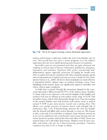
Fig. 7.18 TCC in the trigone showing a classic fimbriated appearance.
surface and forming a solid mass within the wall of the bladder may be
seen. This second type may carry a worse prognosis as in the author’s
experience they are more rapidly growing and refractory to treatment.
Invariably cases are not presented until they are quite advanced and
resulting in clinical signs of urinary obstruction: pollakiuria, stranguria,
haematuria and tenesmus. Treatment with certain non-steroidal anti-
inflammatory agents, especially piroxicam (0.3 mg/kg by mouth every
24 h), is useful and may be combined with other chemotherapeutic agents
such as mitoxantrone (5 mg/m2 intravenous every 3 weeks for four treat-
ments) (Upton et al., 2006). However these treatments are most effective
if introduced before clinical signs of urethral blockage occur. Laser
debulking of the tumour tissue can significantly improve morbidity and
relieve clinical signs completely.
A diode laser is passed through the instrument channel of the cysto-
scope and used to ablate around 90–95% of the tumour tissue. Neoplas-
tic tissue which is not adjacent to the normal margins of the bladder and
urethral wall may be ablated in non-contact mode using 10–15 W. This
devitalises tissue, which will subsequently slough away. Near the margins
of the normal bladder wall and urethral wall contact mode is used at
around 9–10 W to give more precise control and a cutting effect. This
allows very careful resection of tumour tissues with minimal risk of
perforation (Fig. 7.19). If haemorrhage obscures the view the bladder
may be drained and filled with carbon dioxide or room air. The former
is safer as the risk of air embolism is reduced. Laser ablation carried out
in air carries additional risk of perforation since tissue heating and col-
lateral spread are greater without the heat-sink effect of fluid. Great care
must be taken not to devitalise deep tissues. Ablation must be done care-
fully and slowly and can be a time-consuming procedure. Extensive
neoplasia may take 2–3 h or longer to ablate.