
Page 233 - Clinical Manual of Small Animal Endosurgery
P. 233
Female Reproductive Tract 221
(a) (b) (c)
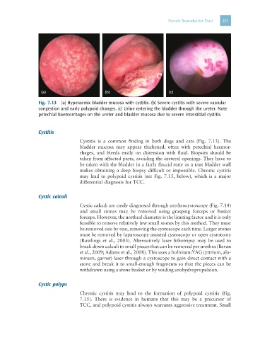
Fig. 7.13 (a) Hyperaemic bladder mucosa with cystitis. (b) Severe cystitis with severe vascular
congestion and early polypoid changes. (c) Urine entering the bladder through the ureter. Note
petechial haemorrhages on the ureter and bladder mucosa due to severe interstitial cystitis.
Cystitis
Cystitis is a common finding in both dogs and cats (Fig. 7.13). The
bladder mucosa may appear thickened, often with petechial haemor-
rhages, and bleeds easily on distension with fluid. Biopsies should be
taken from affected parts, avoiding the ureteral openings. They have to
be taken with the bladder in a fairly flaccid state as a taut bladder wall
makes obtaining a deep biopsy difficult or impossible. Chronic cystitis
may lead to polypoid cystitis (see Fig. 7.15, below), which is a major
differential diagnosis for TCC.
Cystic calculi
Cystic calculi are easily diagnosed through urethrocystoscopy (Fig. 7.14)
and small stones may be removed using grasping forceps or basket
forceps. However, the urethral diameter is the limiting factor and it is only
feasible to remove relatively few small stones by this method. They must
be removed one by one, removing the cystoscope each time. Larger stones
must be removed by laparoscopy-assisted cystoscopy or open cystotomy
(Rawlings et al., 2003). Alternatively laser lithotripsy may be used to
break down calculi to small pieces that can be removed per urethra (Bevan
et al., 2009; Adams et al., 2008). This uses a holmium:YAG (yttrium, alu-
minum, garnet) laser through a cystoscope to gain direct contact with a
stone and break it to small-enough fragments so that the pieces can be
withdrawn using a stone basket or by voiding urohydropropulsion.
Cystic polyps
Chronic cystitis may lead to the formation of polypoid cystitis (Fig.
7.15). There is evidence in humans that this may be a precursor of
TCC, and polypoid cystitis always warrants aggressive treatment. Small