
Page 228 - Clinical Manual of Small Animal Endosurgery
P. 228
216 Clinical Manual of Small Animal Endosurgery
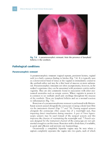
Fig. 7.6 A paramesonephric remnant. Note the presence of lymphoid
follicles in the vestibule.
Pathological conditions
Paramesonephric remnant
A paramesonephric remnant (vaginal septum, persistent hymen, vaginal
web) is a fairly common finding in bitches (Fig. 7.6). It is typically seen
as a dorsoventral band of tissue at the vaginal os immediately cranial to
the urethral orifice and may be a thin band of mucosa or more substan-
tial. Paramesonephric remnants are often asymptomatic; however, in the
author’s experience they can be associated with persistent cystitis and/or
vaginitis. They are also commonly found in association with other ana-
tomical anomalies such as ectopic ureters. Where vaginitis is present it
is common to see multiple small pale swellings throughout the mucosa
of the vestibule. These are lymphoid follicles and are a normal reaction
to inflammation (Fig. 7.6).
Resection of a paramesonophrenic remnant is performed with Metzen-
baum scissors passed alongside the cystoscope or using a diode laser fibre
via the instrument channel (Figs 7.7 and 7.8). Passing surgical scissors
alongside the cystoscope often causes loss of a watertight seal, thus
impairing direct visualisation during resection. A pair of 5 mm laparo-
scopic scissors may be used instead of the surgical scissors and this
improves the chances of maintaining the watertight seal. 7 French scis-
sors designed for the instrument channel of the cystoscope are not sub-
stantial enough to cut this tissue. Resection with a diode laser, if available,
is quick and efficient and prevents any haemorrhage.
Occasionally a completely bipartite vagina may be seen where a
septum completely separates the vagina into two parts, each of which