
Page 226 - Clinical Manual of Small Animal Endosurgery
P. 226
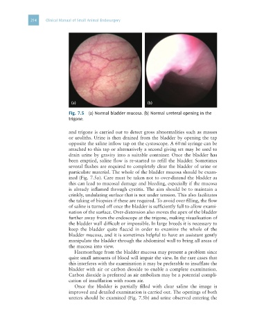
214   Clinical Manual of Small Animal Endosurgery


                               (a)                             (b)

                              Fig. 7.5  (a) Normal bladder mucosa. (b) Normal ureteral opening in the
                              trigone.


                              and trigone is carried out to detect gross abnormalities such as masses
                              or uroliths. Urine is then drained from the bladder by opening the tap
                              opposite the saline inflow tap on the cystoscope. A 60 ml syringe can be
                              attached to this tap or alternatively a second giving set may be used to
                              drain urine by gravity into a suitable container. Once the bladder has
                              been emptied, saline flow is re-started to refill the bladder. Sometimes
                              several flushes are required to completely clear the bladder of urine or
                              particulate material. The whole of the bladder mucosa should be exam-
                              ined (Fig. 7.5a). Care must be taken not to over-distend the bladder as
                              this can lead to mucosal damage and bleeding, especially if the mucosa
                              is already inflamed through cystitis. The aim should be to maintain a
                              crinkly, undulating surface that is not under tension. This also facilitates
                              the taking of biopsies if these are required. To avoid over-filling, the flow
                              of saline is turned off once the bladder is sufficiently full to allow exami-
                              nation of the surface. Over-distension also moves the apex of the bladder
                              further away from the endoscope at the trigone, making visualisation of
                              the bladder wall difficult or impossible. In large breeds it is necessary to
                              keep  the  bladder  quite  flaccid  in  order  to  examine  the  whole  of  the
                              bladder mucosa, and it is sometimes helpful to have an assistant gently
                              manipulate the bladder through the abdominal wall to bring all areas of
                              the mucosa into view.
                                Haemorrhage from the bladder mucosa may present a problem since
                              quite small amounts of blood will impair the view. In the rare cases that
                              this interferes with the examination it may be preferable to insufflate the
                              bladder with air or carbon dioxide to enable a complete examination.
                              Carbon dioxide is preferred as air embolism may be a potential compli-
                              cation of insufflation with room air.
                                Once  the  bladder  is  partially  filled  with  clear  saline  the  image  is
                              improved and detailed examination is carried out. The openings of both
                              ureters should be examined (Fig. 7.5b) and urine observed entering the