
Page 225 - Clinical Manual of Small Animal Endosurgery
P. 225
Female Reproductive Tract 213
appearance of the vaginal wall will vary according to the stage of the
oestrus cycle. The walls become thickened and more folded during
oestrus, becoming thinner and less prominent during anoestrus.
Having examined the vagina, the cystoscope is withdrawn as far as
the vestibule and redirected ventrally into the urethral opening. Saline
flow is turned on again to distend the urethra as the cystoscope passes
down. Once in the urethra, the surgeon may relax the grip on the vulva
as a watertight seal is no longer required. Great care must be taken when
passing a 30° endoscope down a narrow lumen such as the urethra. The
natural instinct is to keep the lumen central in the field of view. However,
with a 30° endoscope this would result in the tip of the endoscope
running along the mucosa at an angle of 30° which could result in
damage or even perforation. Viewing the image as you pass a bare endo-
scope into the cystoscopy sheath will demonstrate that the lumen of the
sheath is right at the bottom of the image on the monitor. This is the
orientation that must be maintained in the urethral lumen. It is standard
for the angle of view of rigid endoscopes to be directed away from the
light post, and the endoscopist can use this to maintain orientation
during the procedure.
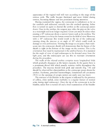
The walls of the relaxed urethra comprise many longitudinal folds
which gradually disappear as the lumen expands. In the queen there is
a prominent dorsal fold which usually remains visible throughout the
procedure. The mucosa should look pink and smooth (Fig. 7.4) and
should be observed for abnormalities as the cystoscope passes down the
urethra. Erythema, petechial haemorrhages, transitional cell carcinoma
(TCC) or the openings of ectopic ureters are easily seen (see later).
The entrance of the bladder at the trigone is indicated by the presence
of yellow, often turbid, urine (turbidity is a regular feature of normal
urine during rigid endoscopy). Once the tip of the cystoscope is in the
bladder, saline flow is turned off and a brief examination of the bladder
Fig. 7.4 A normal urethra.