
Page 223 - Clinical Manual of Small Animal Endosurgery
P. 223
Female Reproductive Tract 211
Urethrocystoscopy procedure
Urethrocystoscopy is performed under general anaesthesia, usually on a
tub table or on a wire grid over a suitable tray for collecting fluid. Urine
samples for bacterial culture are indicated in most candidates for ure-
throcystoscopy and are best obtained by cystocentesis prior to the pro-
cedure. The room is arranged so that the monitor is at the head of the
patient, which makes orientation during the examination more straight-
forward. The patient may be positioned in ventral, lateral or dorsal
recumbency according to personal preference. For routine urethrocystos-
copy this author prefers to position the patient in ventral recumbency
with a rolled up towel under the caudal abdomen to elevate the pelvis a
little. The tail is held or taped out of the way. In this position anatomical
structures are in their normal orientation and a full examination can be
carried out following a set routine.
The endoscope is mounted into the cystoscope sheath and the camera
and light guide cable attached. The light source is switched on and the
camera white-balanced. A litre bag of sterile saline is attached via a
giving set to one of the Luer taps on the cystoscope sheath and the clamps
on the giving set are opened so that flow can be controlled with the
forefinger operating the Luer tap on the cystoscope sheath.
It is not usually necessary to clip the peri-vulvar hair unless it is very
long or is soiled and matted. The cystoscope sheath is lubricated with a
little sterile water-soluble lubricating gel and the tip is introduced into
the dorsal commisure of the vulva in a cranio-dorsal direction to avoid
the clitoral fossa. With the tip of the cystoscope just inside the vulvar
lips, firm pressure is applied with the thumb and forefinger to form a
watertight seal around the sheath. The saline flow is turned on and the
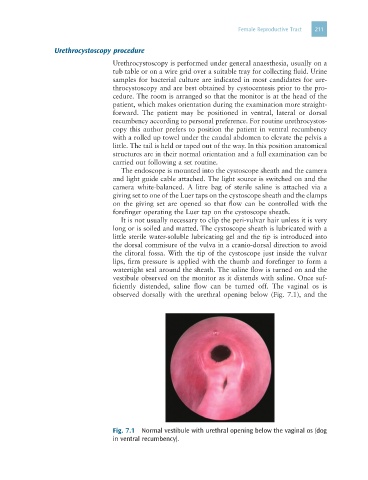
vestibule observed on the monitor as it distends with saline. Once suf-
ficiently distended, saline flow can be turned off. The vaginal os is
observed dorsally with the urethral opening below (Fig. 7.1), and the
Fig. 7.1 Normal vestibule with urethral opening below the vaginal os (dog
in ventral recumbency).