
Page 224 - Clinical Manual of Small Animal Endosurgery
P. 224
212 Clinical Manual of Small Animal Endosurgery
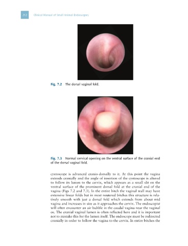
Fig. 7.2 The dorsal vaginal fold.
Fig. 7.3 Normal cervical opening on the ventral surface of the cranial end
of the dorsal vaginal fold.
cystoscope is advanced cranio-dorsally to it. At this point the vagina
extends cranially and the angle of insertion of the cystoscope is altered
to follow its lumen to the cervix, which appears as a small slit on the
ventral surface of the prominent dorsal fold at the cranial end of the
vagina (Figs 7.2 and 7.3). In the entire bitch the vaginal wall may have
extensive linear folds but in most neutered bitches this structure is rela-
tively smooth with just a dorsal fold which extends from about mid
vagina and increases in size as it approaches the cervix. The endoscopist
will often encounter an air bubble in the caudal vagina near the vaginal
os. The cranial vaginal lumen is often reflected here and it is important
not to mistake this for the lumen itself. The endoscope must be redirected
cranially in order to follow the vagina to the cervix. In entire bitches the