
Page 234 - Clinical Manual of Small Animal Endosurgery
P. 234
222 Clinical Manual of Small Animal Endosurgery
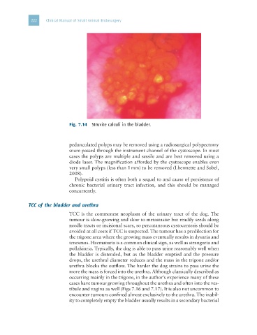
Fig. 7.14 Struvite calculi in the bladder.
pedunculated polyps may be removed using a radiosurgical polypectomy
snare passed through the instrument channel of the cystoscope. In most
cases the polyps are multiple and sessile and are best removed using a
diode laser. The magnification afforded by the cystoscope enables even
very small polyps (less than 1 mm) to be removed (Lhermette and Sobel,
2008).
Polypoid cystitis is often both a sequel to and cause of persistence of
chronic bacterial urinary tract infection, and this should be managed
concurrently.
TCC of the bladder and urethra
TCC is the commonest neoplasm of the urinary tract of the dog. The
tumour is slow-growing and slow to metastasise but readily seeds along
needle tracts or incisional scars, so percutaneous cystocentesis should be
avoided at all costs if TCC is suspected. The tumour has a predilection for
the trigone area where the growing mass eventually results in dysuria and
tenesmus. Haematuria is a common clinical sign, as well as stranguria and
pollakiuria. Typically, the dog is able to pass urine reasonably well when
the bladder is distended, but as the bladder emptied and the pressure
drops, the urethral diameter reduces and the mass in the trigone and/or
urethra blocks the outflow. The harder the dog strains to pass urine the
more the mass is forced into the urethra. Although classically described as
occurring mainly in the trigone, in the author’s experience many of these
cases have tumour growing throughout the urethra and often into the ves-
tibule and vagina as well (Figs 7.16 and 7.17). It is also not uncommon to
encounter tumours confined almost exclusively to the urethra. The inabil-
ity to completely empty the bladder usually results in a secondary bacterial