
Page 53 - Live-cellanalysis handbook
P. 53
Kinetic Scratch Wound Assays
Two-color fluorescent scratch wound experiments
In addition to phase contrast images, two-color images can be with IncuCyte® NucLight Green Lentivirus Reagent, and the
collected using IncuCyte® fluorescent labeling reagents. This HT-1080 cells labeled with IncuCyte® NucLight Red Lentivirus
allows study of the interactions between several cell types in a Reagent were mixed in co-culture, and plated for an invasion
mixed culture, and how each affects migration, invasion, and assay through 8 mg/ml Matrigel®. Imaging in phase, red, and
proliferation of the other, all within one well of a 96-well plate. green channels revealed the HT- 1080 cells efficiently invaded
As shown in Figure 5, the non-invasive MCF- 7 cells were labeled the Matrigel® matrix whereas MCF-7 remained non-invasive.
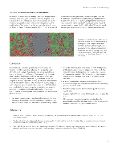
Figure 5. Two color fluorescent scratch wound.
The ability to image cells in both wavelengths
in addition to phase contrast allows users
the ability to explore cell-cell interactions
as it pertains to cell migration and invasion.
In this example, HT-1080 NucLight Red cells
were plated with MCF-7 NucLight Green cells
and invasion through 8 mg/ml Matrigel® was
monitored over time (Image shows the 24 hour
time point).
Conclusions
IncuCyte scratch wound migration and invasion assays are • The spatio-temporal, label-free format of both the Migration
flexible, quantitative, and reproducible. The unique tip design and Invasion Assays allows investigators to follow both the
of the 96-well IncuCyte WoundMaker is a critical part of these rate and the extent of migration and invasion for a given set
assays, as it creates a cell-free zone from a confluent monolayer of experimental variables. This feature can be used to explore
of cells making the biology consistent and reproducible. Both time-dependent pharmacology, in order to enhance assay
assays utilize IncuCyte’s innovative high definition optics and sensitivity.
integrative analysis algorithms to make quantitative measurements • After the experiment is initiated, phase contrast and/ or
without the need for labeling cells. Having high-quality images at fluorescence images are collected and processed automatically,
every time point gives an investigator access to both the kinetics yielding un-biased results.
and morphological changes occurring in migration and invasion
experiments, enabling additional insight intothe effects test • Precise wounding ensures that results are quantitative and
agents have on cells undergoing these processes. Key features and reproducible.
benefits include: • IncuCyte high definition optics eliminates the need to label the
cells.
• Investigators can measure migration and invasion on the same • HD images are acquired at every time point and can be
microplate. This provides the best opportunity for determining automatically assembled into time-lapse movies for convenient
the specificity of drugs and the utility of potential drug targets.
visualization of morphology and wound closure.
References
1. Kovacs, M., Toth, J,, Hetnyi, C., Malnasi-Csizmadia, A and Sellers, J.R. Mechanism of action of Blebbistatin inhibition of Myosin II. J. Biol. Chem.
279(34) 35557 (2004)
2. Poinclouxa,R., Collina, O., Lizárragaa F., Romaoa, M., Debraye, M., Piela, M. and Chavrier, P. Contractility of the cell rear drives invasion of breast
tumor cells in 3D Matrigel. PNAS 108 (5):1943 (2011)
3. Fischer, K.E., Pop, A., Koh, W., Anthis, N.J., Saunders,W.B. and Davis, G.E. Tumor cell invasion of collagen matrices requires coordinate lipid agonist-
induced G-protein and membrane-type matrix metalloproteinase-1-dependent signaling. Mol Cancer. 5:69 (2006)
51