
Page 213 - Fluid, Electrolyte, and Acid-Base Disorders in Small Animal Practice
P. 213
Disorders of Phosphorus: Hypophosphatemia and Hyperphosphatemia 203
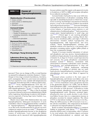
BOX 7-2 Causes of because acidosis caused by organic acids apparently results
in breakdown of ATP to AMP and inorganic phosphate
Hyperphosphatemia by an unknown mechanism. 119
Increased intake of phosphorus may occur with intra-
Maldistribution (Translocation) venous administration of phosphate-containing fluids,
• Tumor cell lysis especially in immobilized patients in which bone resorp-
• Tissue trauma or rhabdomyolysis tion is occurring. Such therapy is uncommon in veterinary
• Hemolysis practice, except in the treatment of diabetic ketoacidosis
• Metabolic acidosis and total parenteral nutrition. 23,173 Increased absorption
Increased Intake of phosphorus from the alimentary tract may occur with
colonic infusion of hypertonic enema solutions or oral
• Gastrointestinal 47
○ Phosphate enemas administration of sodium phosphate. Such enemas have
○ Vitamin D intoxication (e.g., cholecalciferol- caused severe hyperphosphatemia in small dogs and
containing rodenticides, calcipotriene) cats. 9,78,147 Clinical signs in cats receiving phosphate
• Parenteral enemas include lethargy, ataxia, vomiting, bloody diar-
○ Intravenous phosphate rhea, mucous membrane pallor, and stupor. Laboratory
Decreased Excretion abnormalities included marked hyperglycemia and
hyperphosphatemia, mild hypernatremia, and lactic
• Acute or chronic renal failure 9
• Uroabdomen or urethral obstruction acidosis. Severe hyperphosphatemia, azotemia, and
• Hypoparathyroidism metabolic acidosis were reported in a cat treated with a
• Acromegaly (?)* phosphate-containing urinary acidifier (pHos-pHaid) at
• Hyperthyroidism twice the recommended dosage. 67
Physiologic: Young Growing Animal Vitamin D increases intestinal absorption of calcium
and phosphorus and may produce hyperphosphatemia
in addition to hypercalcemia. In one study, administration
Laboratory Error (e.g., Lipemia, of vitamin D 2 to dogs for 3 weeks caused hypercalcemia
Hyperproteinemia) Depending on and azotemia, but serum phosphorus concentrations
Methodology remained normal. 165 However, intoxication with
cholecalciferol-containing rodenticides causes azotemia,
hypercalcemia, and hyperphosphatemia in dogs and
*(?) Importance in veterinary medicine uncertain. 48,60,69,104,114
cats. Topical medications containing
calcipotriene, an analogue of calcitriol, also can cause
hypercalcemia, hyperphosphatemia, metastatic soft tissue
increased. There was no change in FE Pi or renal function mineralization, and acute renal failure if ingested by
(as assessed by endogenous creatinine clearance). Chemo- dogs. 51,71,124
therapy in these dogs consisted of prednisone, vincristine, Decreased urinary excretion is the main cause of
and L-asparaginase. However, acute tumor lysis syndrome hyperphosphatemia, and chronic renal failure is the most
has been reported in some animals with lymphosarcoma common cause of hyperphosphatemia in adult dogs and
treated with chemotherapy with or without radiation ther- cats. 31 Chronic renal disease causes a progressive decrease
apy. 28,93,94 Severe hyperphosphatemia (23.6 and 13.7 in the glomerular filtration rate (GFR), and the filtered
mg/dL) occurred in a dog and a cat (respectively), and load of phosphate (GFR serum phosphorus concentra-
mild hyperphosphatemia (7.4 and 7.7 mg/dL) occurred tion) decreases as GFR decreases. If phosphorus intake
in two other affected dogs. 28,93 Thus, it may be prudent remains constant, phosphorus retention and transient
to promote diuresis by intravenous administration of fluids hyperphosphatemia result. However, sustained hyper-
before beginning chemotherapy in patients with lympho- phosphatemia does not usually develop in early chronic
sarcoma suspected of having large tumor burdens (e.g., renal failure because there is a compensatory increase in
hepatosplenomegaly). phosphate excretion by remnant nephrons. The effects
Massive tissue injury with rhabdomyolysis may cause of PTH on the kidneys mediate this increase in the FE Pi .
hyperphosphatemia. Subsequent development of acute When the GFR decreases to 20% of normal or less (i.e.,
renal failure related to myoglobinuria further contributes late chronic renal failure), this compensatory mechanism
to hyperphosphatemia. 164 Hyperphosphatemia may is exhausted and hyperphosphatemia develops.
occur after aortic thromboembolism in cats and was more Renal secondary hyperparathyroidism is a consistent
common in nonsurvivors in one study. 163 Hemolysis can finding in progressive renal disease. 158,160 Hyperphos-
produce hyperphosphatemia because of the phosphorus phatemia inhibits renal 1a-hydroxylase, which is present
content of erythrocytes. Lactic acidosis and diabetic in the renal tubules (this inhibition impairs conversion
ketoacidosis can be associated with hyperphosphatemia of 25-hydroxycholecalciferol to calcitriol and thus