Page 209 - Feline Cardiology
P. 209
214  Section F: Arrhythmias and Other Electrocardiographic Abnormalities


                                                                   reverse the cause of the bradycardia (e.g., reduce the
                                                                   concentration of inhalant anesthetic), and if not suf-
                                                                   ficient,  treatment  to  increase  the  heart  rate  directly
                Cardiac Output (ml/min)                            (e.g., glycopyrrolate for sinus bradycardia under anes-
                                                                   thesia, or pacemaker implantation for AV block and a
                                                                   low ventricular escape rate) are essential in order to
                                                                   promptly  raise  the  heart  rate  and  return  the  cardiac
                                                                   output to a level compatible with life.
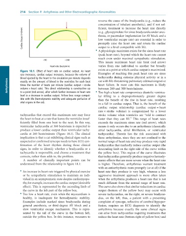
                                                                 •  A physiologic maximum exists for the sinus heart rate
                                                                   (peak heart rate), beyond which the heart rate cannot
                                                                   reach  even  under  maximal  sympathetic  stimulation.
                              ≈90           ≈280
                                                                   This  innate  maximum  heart  rate  limit  (red  arrow)
                                  Heart rate (beats/min)           varies  from  one  individual  to  another  but  broadly
                                                                   exists at a point at which cardiac output is maximized.
               Figure  18.1.  Effect	 of	 heart	 rate	 on	 cardiac	 output.	 As	 heart
               rate	increases,	cardiac	output	increases,	because	the	volume	of	  Examples of reaching this peak heart rate are sinus
               blood	ejected	by	the	heart	to	the	circulation	per	minute	depends	  tachycardia during extreme physical activity or in a
      Arrhythmias  times	the	number	of	beats	per	minute	(cardiac	output	=	stroke	  heart  failure.  In  most  cats  this  maximum  is  likely
                                                                   cat with life-threatening pulmonary edema/congestive
               exactly	on	the	amount	of	blood	in	the	ventricles	for	each	beat
                                                                   between 260 and 300 beats/minute.
               volume	×	heart	rate).	This	direct	relationship	is	constructive	up
               to	a	point	(red	arrow),	after	which	further	increases	in	heart	rate
                                                                   lar  filling  to  a  disproportionately  greater  degree
               lead	to	a	decrease	in	cardiac	output.	Yellow	box:	range	compat-  •  Too high a heart rate compromises diastolic ventricu-
               ible	with	life	(hemodynamic	stability	and	adequate	perfusion	of	  than  the  benefit  of  the  rise  in  heart  rate,  resulting
               vital	organs	in	the	cat).                           in a fall in cardiac output. That is, the benefit of the
                                                                   cardiac  output  relationship  (cardiac  output = heart
                                                                   rate × stroke  volume)  is  compromised  by  a  lower
              tachycardias that exceed this maximum rate may force   stroke volume when ventricles are “told to contract
              the heart to beat at a rate that leaves the ventricles insuf-  faster  than  they  can  fill.”  This  range  of  heart  rates
              ficiently  filled  from  one  beat  to  the  next.  In  this  way,   exceeds the maximum sinus rate (red arrow), which
              ventricular tachycardia at 360 beats/min is expected to   means it only occurs due to an arrhythmia (classically
              produce a lower cardiac output than ventricular tachy-  atrial  tachycardia,  atrial  fibrillation,  or  ventricular
              cardia  at  260  beats/minute  (Figure  18.1).  The  clinical   tachycardia).  Therein  lies  the  risk  associated  with
              implication is that a cat exhibiting clinical signs such as   these arrhythmias, since they are not confined to the
              suspected or confirmed syncope needs to have ECG con-  normal range of heart rate and may produce very rapid
              firmation  of  the  heart  rhythm  during  those  clinical   tachycardias that markedly reduce cardiac output (the
              signs,  in  order  to  identify  whether  a  bradycardia  or  a   descending limb on the right side of the curve within
              tachycardia is responsible, and choose a treatment that   the yellow box). This region of the curve illustrates
              corrects, rather than adds to, the problem.          that tachycardias generally produce negative hemody-
                 A  number  of  clinically  important  points  can  be   namic effects that are more severe when the heart rate
              understood from the relationship in figure 18.1:     is  higher.  Therefore,  arrhythmias  warrant  treatment
                                                                   with an antiarrhythmic more promptly (or at all) if the
              •  An increase in heart rate triggered by physical exercise   heart rate they produce is very high, whereas a less
                or  by  sympathetic  stimulation  to  maintain  an  indi-  aggressive  treatment  approach  is  more  often  taken
                vidual in an asymptomatic state despite cardiomyopa-  when the arrhythmia produces a heart rate that is not
                thy, for example, increases the cardiac output (desirable   much different from the normal range of heart rate.
                effect). This is represented by the ascending limb of   The curve also shows that similar reductions in cardiac
                the curve in the left part of the yellow box.      output  (bottom  of  the  yellow  box)  may  occur  with
              •  Too low a heart rate, even when the myocardium is   severe tachycardias, on the right, or severe bradycar-
                healthy,  is  inadequate  for  hemodynamic  stability.   dias,  on  the  left;  thus,  a  patient  with  a  presenting
                Examples  include  marked  sinus  bradycardia  during   complaint of syncope, reflective of cerebral hypoper-
                general  anesthesia,  or  third-degree AV  block  and  a   fusion,  requires  an  ECG  diagnosis  to  identify  the
                slow  ventricular  escape  mechanism.  This  is  repre-  arrhythmia  because  exactly  the  same  clinical  signs
                sented  by  the  tail  of  the  curve  in  the  bottom  left,   can arise from tachycardias requiring treatments that
                outside the yellow box. In this instance, measures to   reduce the heart rate (bottom right of yellow box) and

