Page 212 - Feline Cardiology
P. 212
Chapter 18: Arrhythmias and Other Electrocardiographic Abnormalities  217


                                                                                                      QRS


                                                                                                    P       T


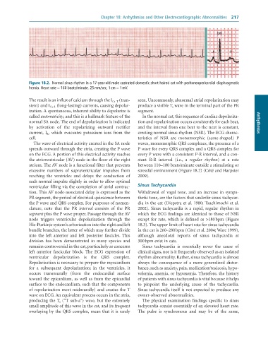
              Figure 18.2.  Normal	sinus	rhythm	in	a	17-year-old	male	castrated	domestic	short-haired	cat	with	peritoneopericardial	diaphragmatic
              hernia.	Heart	rate	=	160	beats/minute.	25	mm/sec,	1	cm	=	1	mV.

              The result is an influx of calcium through the I Ca–T  (tran-  seen. Uncommonly, abnormal atrial repolarization may
              sient) and I Ca–L  (long-lasting) currents, causing depolar-  produce a visible T a  wave in the terminal part of the PR
              ization. A spontaneous, inherent ability to depolarize is   segment.
              called automaticity, and this is a hallmark feature of the   In the normal cat, this sequence of cardiac depolariza-
              normal SA node. The end of depolarization is indicated   tion and repolarization occurs consistently for each beat,   Arrhythmias
              by  activation  of  the  repolarizing  outward  rectifier   and the interval from one beat to the next is constant,
              current,  I K,  which  evacuates  potassium  ions  from  the   creating normal sinus rhythm (NSR). The ECG charac-
              cell.                                              teristics  of  NSR  are  monomorphic  (same-shaped)  P
                 The wave of electrical activity created in the SA node   waves, monomorphic QRS complexes, the presence of a
              spreads outward through the atria, creating the P wave   P wave for every QRS complex and a QRS complex for
              on the ECG. A portion of this electrical activity reaches   every P wave with a consistent P-R interval, and a con-
              the atrioventricular (AV) node in the floor of the right   stant  R-R  interval  (i.e.,  a  regular  rhythm)  at  a  rate
              atrium. The AV node is a functional filter that prevents   between 110–180 beats/minute outside a stimulating or
              excessive  numbers  of  supraventricular  impulses  from   stressful environment (Figure 18.2) (Côté and Harpster
              reaching  the  ventricles  and  delays  the  conduction  of   2009).
              each normal impulse slightly in order to allow optimal
              ventricular filling via the completion of atrial contrac-  Sinus Tachycardia
              tion. This AV node-associated delay is expressed as the   Withdrawal  of  vagal  tone,  and  an  increase  in  sympa-
              PR segment, the period of electrical quiescence between   thetic tone, are the factors that underlie sinus tachycar-
              the P wave and QRS complex. For purposes of nomen-  dia in the cat (Diepstra et al. 1980; Tsuchimochi et al.
              clature,  note  that  the  PR  interval  consists  of  the  PR   2002). Sinus tachycardia is a rapid, regular rhythm in
              segment plus the P wave proper. Passage through the AV   which the ECG findings are identical to those of NSR
              node  triggers  ventricular  depolarization  through  the   except  for  rate,  which  is  defined  as  >180 bpm  (Figure
              His-Purkinje system’s arborization into the right and left   18.3). The upper limit of heart rate for sinus tachycardia
              bundle branches, the latter of which may further divide   in the cat is 260–280 bpm (Côté et al. 2004; Ware 1999),
              into  the  left  anterior  and  left  posterior  fascicles.  This   although  anecdotal  reports  of  sinus  tachycardia  at
              division  has  been  demonstrated  in  many  species  and   300 bpm exist in cats.
              remains controversial in the cat, particularly as concerns   Sinus  tachycardia  is  essentially  never  the  cause  of
              left  anterior  fascicular  block.  The  ECG  expression  of   clinical signs, nor is it frequently observed as an isolated
              ventricular  depolarization  is  the  QRS  complex.   rhythm abnormality. Rather, sinus tachycardia is almost
              Repolarization is necessary to prepare the myocardium   always  the  consequence  of  a  more  generalized  distur-
              for  a  subsequent  depolarization;  in  the  ventricles,  it   bance, such as anxiety, pain, medication/toxicosis, hypo-
              occurs  transmurally  (from  the  endocardial  surface   volemia, anemia, or hypoxemia. Therefore, the history
              toward  the  epicardium,  as  well  as  from  the  epicardial   of patients with sinus tachycardia is vital because it helps
              surface to the endocardium, such that the components   to  pinpoint  the  underlying  cause  of  the  tachycardia.
              of repolarization meet midmurally) and creates the T   Sinus tachycardia itself is not expected to produce any
              wave on ECG. An equivalent process occurs in the atria,   owner-observed abnormalities.
              producing the T a  (“T sub-a”) wave, but the extremely   The  physical  examination  findings  specific  to  sinus
              small amplitude of this wave in the cat, and its frequent   tachycardia consist essentially of an elevated heart rate.
              overlaying by the QRS complex, mean that it is rarely   The  pulse  is  synchronous  and  may  be  of  the  same,

