Page 215 - Feline Cardiology
P. 215
220  Section F: Arrhythmias and Other Electrocardiographic Abnormalities


                                                                                                   P
                                                                                                           T


                                                                                                     QRS


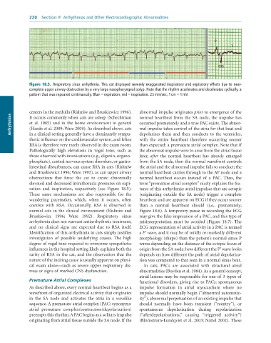
              Figure 18.5.  Respiratory	sinus	arrhythmia.	This	cat	displayed	severely	exaggerated	inspiratory	and	expiratory	efforts	due	to	near-
              complete	upper	airway	obstruction	by	a	very	large	nasopharyngeal	polyp.	Note	that	the	rhythm	accelerates	and	decelerates	cyclically,	a
              pattern	that	was	repeated	continuously.	Blue	=	expiration;	red	=	inspiration.	25	mm/sec,	1	cm	=	1	mV.


              centers in the medulla (Rishniw and Bruskiewicz 1996).   abnormal impulse originates prior to emergence of the
      Arrhythmias  et  al.  1985)  and  in  the  home  environment  in  general   occurred prematurely and a true PAC exists. The abnor-
              It occurs commonly when cats are asleep (Schechtman
                                                                 normal  heartbeat  from  the  SA  node,  the  impulse  has
                                                                 mal impulse takes control of the atria for that beat and
              (Hanås et al. 2009; Ware 2009). As described above, cats
              in a clinical setting generally have a dominantly sympa-
                                                                 with  the  entire  heartbeat  therefore  occurring  sooner
              thetic influence on the cardiovascular system, and feline   depolarizes  them  and  then  conducts  to  the  ventricles,
              RSA is therefore very rarely observed in the exam room.   than expected: a premature atrial complex. Note that if
              Pathologically  high  elevations  in  vagal  tone,  such  as   the abnormal impulse were to arise from the atrial tissue
              those observed with intoxications (e.g., digoxin, organo-  later,  after  the  normal  heartbeat  has  already  emerged
              phosphate), central nervous system disorders, or gastro-  from the SA node, then the normal wavefront controls
              intestinal disturbances, can cause RSA in cats (Rishniw   the atrial and the abnormal impulse fails to conduct; the
              and Bruskiewicz 1996; Ware 1992), as can upper airway   normal heartbeat carries through to the AV node and a
              obstructions  that  force  the  cat  to  create  abnormally   normal  heartbeat  occurs  instead  of  a  PAC.  Thus,  the
              elevated and decreased intrathoracic pressures on expi-  term “premature atrial complex” nicely explains the fea-
              ration  and  inspiration,  respectively  (see  Figure  18.5).   tures of this arrhythmia: atrial impulses that are ectopic
              These  same  mechanisms  are  also  responsible  for  the   (originating  outside  the  SA  node)  trigger  a  complete
              wandering  pacemaker,  which,  when  it  occurs,  often   heartbeat and are apparent on ECG if they occur sooner
              coexists  with  RSA.  Occasionally,  RSA  is  observed  in   than  a  normal  heartbeat  should  (i.e.,  prematurely;
              normal cats in the clinical environment (Rishniw and   Figure 18.6). A temporary pause in recording the ECG
              Bruskiewicz  1996;  Ware  1992).  Respiratory  sinus   may give the false impression of a PAC, and this type of
              arrhythmia does not warrant antiarrhythmic treatment,   misinterpretation  must  be  avoided  (Figure  18.7).  The
              and  no  clinical  signs  are  expected  due  to  RSA  itself.   ECG representation of atrial activity in a PAC is termed
              Identification of this arrhythmia in cats simply justifies   a P’ wave, and it may be of mildly or markedly different
              investigation  of  possible  underlying  causes.  The  high   morphology (shape) than the patient’s normal sinus P
              degree of vagal tone required to overcome sympathetic   waves depending on the distance of the ectopic focus of
              influences in the hospital setting likely explains both the   origin from the SA node: how different the P’ wave looks
              rarity of RSA in the cat, and the observation that the   depends on how different the path of atrial depolariza-
              nature of the inciting cause is usually apparent on physi-  tion was compared to that seen in a normal sinus beat.
              cal exam alone—such as severe upper respiratory dis-  In  cats,  PACs  are  associated  with  structural  atrial
              tress or signs of marked CNS dysfunction.          abnormalities (Boyden et al. 1984). As a general concept,
                                                                 atrial lesions may be responsible for one of 3 types of
              Premature Atrial Complexes                         functional disorders, giving rise to PACs: spontaneous
              As described above, every normal heartbeat begins as a   impulse  formation  in  atrial  myocardium  where  no
              wavefront of organized electrical activity that originates   impulse should normally begin (“abnormal automatic-
              in  the  SA  node  and  activates  the  atria  in  a  wavelike   ity”), abnormal perpetuation of an existing impulse that
              sequence. A premature atrial complex (PAC; synonyms:   should  normally  have  been  transient  (“reentry”),  or
              atrial  premature  complex/contraction/depolarization)   spontaneous  depolarization  during  repolarization
              preempts this rhythm. A PAC begins as a solitary impulse   (“afterdepolarizations,”  causing  “triggered  activity”)
              originating from atrial tissue outside the SA node. If the   (Blömstrom-Lundqvist et al. 2003; Nattel 2002). These

