Page 216 - Feline Cardiology
P. 216
Chapter 18: Arrhythmias and Other Electrocardiographic Abnormalities  221


                     II
                                                                                        QRS
                                                                                              QRS        QRS
                            *                                  * *
                                                                                    T                  P      T
                                                                                       P     P′    T


               A


                                                                                                    * T   P


                                                                                                           QRS          Arrhythmias

               B
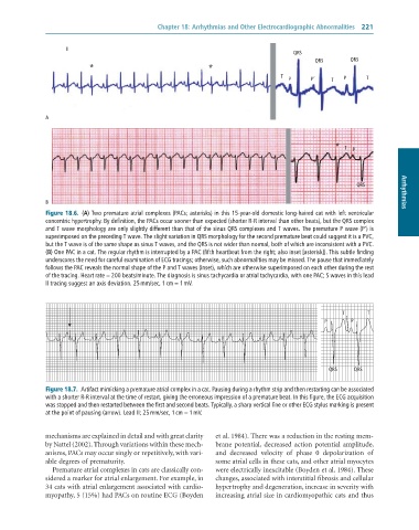
               Figure 18.6.  (A)	Two	premature	atrial	complexes	(PACs;	asterisks)	in	this	15-year-old	domestic	long-haired	cat	with	left	ventricular
               concentric	hypertrophy.	By	definition,	the	PACs	occur	sooner	than	expected	(shorter	R-R	interval	than	other	beats),	but	the	QRS	complex
               and	T	wave	morphology	are	only	slightly	different	than	that	of	the	sinus	QRS	complexes	and	T	waves.	The	premature	P	wave	(P’)	is
               superimposed	on	the	preceding	T	wave.	The	slight	variation	in	QRS	morphology	for	the	second	premature	beat	could	suggest	it	is	a	PVC,
               but	the	T	wave	is	of	the	same	shape	as	sinus	T	waves,	and	the	QRS	is	not	wider	than	normal,	both	of	which	are	inconsistent	with	a	PVC.
               (B)	One	PAC	in	a	cat.	The	regular	rhythm	is	interrupted	by	a	PAC	(fifth	heartbeat	from	the	right;	also	inset	[asterisk]).	This	subtle	finding
               underscores	the	need	for	careful	examination	of	ECG	tracings;	otherwise,	such	abnormalities	may	be	missed.	The	pause	that	immediately
               follows	the	PAC	reveals	the	normal	shape	of	the	P	and	T	waves	(inset),	which	are	otherwise	superimposed	on	each	other	during	the	rest
               of	the	tracing.	Heart	rate	=	200	beats/minute.	The	diagnosis	is	sinus	tachycardia	or	atrial	tachycardia,	with	one	PAC;	S	waves	in	this	lead
               II	tracing	suggest	an	axis	deviation.	25	mm/sec,	1	cm	=	1	mV.


                                                                                                      T       T
                                                                                                 P       P


                                                                                                  QRS     QRS

               Figure 18.7.  Artifact	mimicking	a	premature	atrial	complex	in	a	cat.	Pausing	during	a	rhythm	strip	and	then	restarting	can	be	associated
               with	a	shorter	R-R	interval	at	the	time	of	restart,	giving	the	erroneous	impression	of	a	premature	beat.	In	this	figure,	the	ECG	acquisition
               was	stopped	and	then	restarted	between	the	first	and	second	beats.	Typically,	a	sharp	vertical	line	or	other	ECG	stylus	marking	is	present
               at	the	point	of	pausing	(arrow).	Lead	II;	25	mm/sec,	1	cm	=	1	mV.


              mechanisms are explained in detail and with great clarity   et al. 1984). There was a reduction in the resting mem-
              by Nattel (2002). Through variations within these mech-  brane  potential,  decreased  action  potential  amplitude,
              anisms, PACs may occur singly or repetitively, with vari-  and  decreased  velocity  of  phase  0  depolarization  of
              able degrees of prematurity.                       some atrial cells in these cats, and other atrial myocytes
                 Premature atrial complexes in cats are classically con-  were electrically inexcitable (Boyden et al. 1984). These
              sidered a marker for atrial enlargement. For example, in   changes, associated with interstitial fibrosis and cellular
              34 cats with atrial enlargement associated with cardio-  hypertrophy and degeneration, increase in severity with
              myopathy, 5 (15%) had PACs on routine ECG (Boyden   increasing atrial size in cardiomyopathic cats and thus

