Page 219 - Feline Cardiology
P. 219
224  Section F: Arrhythmias and Other Electrocardiographic Abnormalities


              suppression of sinus tachycardia could cause hemody-  (regular formulation) 7.5 mg PO q 8h, or sotalol 2 mg/kg
              namic collapse; 2) with AT confirmed, determine whether   PO  q  12h.  Sustained-release  diltiazem  (Cardizem-CD,
              AT  alone  appears  responsible  for  the  unstable  state,   Dilacor-XL) is not recommended in cats due to unpre-
              which mainly depends on the heart rate (is it >260 beats/  dictable  bioavailability  and  toxicosis  (Johnson  et  al.
              minute?). If so, identify and correct systemic abnormali-  1996; Wall et al. 2005).
              ties  that  increase  sympathetic  tone  (hypovolemia,   With either acute (IV) or chronic (PO) treatment, the
              anemia, hypoxemia, etc.); 3) in an unstable cat with con-  therapeutic goal is to reduce or eliminate extremes of
              firmed AT at a rate >260/minute and in whom extracar-  tachycardia that would reduce cardiac output—i.e., to
              diac  disorders  are  controlled  or  absent,  intravenous   provide medication at a level that prevents HR >260 and
              diltiazem (0.125 mg/kg IV infusion over 1 minute), pro-  stops  AT-associated  syncope  or  other  arrhythmia-
              pranolol (0.1 mg total bolus/cat, once), or esmolol 25–  associated  clinical  signs  if  present.  Pharmacologically
              200 mcg/kg/min  intravenous  infusion  (begin  low  and   forcing the heart rate beyond these parameters and into
              gradually  increase  to  achieve  desired  effect)  may  be   the  range  of  normal  sinus  rhythm  (110–180  beats/
              administered  with  continuous  ECG  monitoring.  The   minute) would likely require excessive dosages of medi-
              time  to  effect  for  any  of  these  drugs  is  typically  <2   cation and is not recommended.
                                                                   Follow-up  is  warranted  after  7–14  days  of  antiar-
              minutes, and a decrease of 5–20% in heart rate is consid-  rhythmic therapy, or sooner if clinical signs worsen or
      Arrhythmias  change, or an improvement, in the cat’s demeanor, pulse   new clinical signs appear. Such follow-up should consist
              ered  a  satisfactory  response,  together  with  either  no
                                                                 of physical exam, 10-lead resting ECG, and repetition of
              strength,  and  other  hemodynamic  parameters.  If  no
              effect  is  seen,  the  dose  can  be  increased  (esmolol)  or
                                                                 malities if they were noted.
              repeated twice in 10 minutes (diltiazem, propranolol). If   laboratory tests as indicated to evaluate ongoing abnor-
              this approach remains ineffective, the diagnosis should   Atrial  tachycardia  does  not  clearly  confer  a  worse
              be  questioned  (repeat  10-lead  ECG,  including  with  a   prognosis,  and  the  prognosis  for  cats  with  AT  is  not
              vagal maneuver) and logistical errors should be sought   distinguishable from the prognosis of its underlying dis-
              and corrected (is the IV catheter patent?) before addi-  orders (cardiomyopathy, hyperthyroidism, others) when
              tional doses are considered. A cat with AT that is stable   AT is absent.
              (conscious,  breathing  comfortably,  fair  or  good  pulse
              strength) at the time of evaluation may be treated with   Atrial Fibrillation
              atenolol 6.25 mg PO q 12h; uptitration to 12.5 mg PO q   Atrial  fibrillation  (AF)  is  said  to  represent  a  state  of
              12h  is  a  robust  increase  that  may  be  considered  after   electrical chaos in the atria. The atria depolarize from
              several  days  if  AT  persists.  This  higher  dosage  has    multiple foci at a rate of several hundred impulses per
              been used by some cardiologists but may be excessive   minute, many or most of which never cross the AV node
              (causing inappetence, lethargy, and weakness) in some   to  the  ventricles.  The  result  is  a  heart  rate  that  varies
              cats, particularly if there is concurrent structural cardiac   from normal to elevated (e.g., 260/minute) and an irreg-
              disease or other illness. Dilated cardiomyopathy is a rela-  ular rhythm due to the patternless selection of impulses
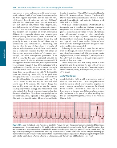
              tive contraindication, especially to the higher dosages.   that are admitted through the AV node to the ventricles
              Alternatives to atenolol include diltiazem hydrochloride   (Figure 18.9).


                                                                                                       QRS


                                                                                                   T         T

              Figure 18.9.  Atrial	fibrillation	in	a	cat.	There	are	no	identifiable	P	waves	(nor	were	they	present	in	any	other	lead);	the	QRS	complexes
              are	monomorphic,	slender,	and	upright	in	this	lead	II	tracing,	and	the	rhythm	is	irregularly	irregular.	The	left	panel	(50	mm/sec)	dem-
              onstrates	that	faster	paper	speeds	show	the	variable	R-R	interval	more	clearly	than	slow	paper	speeds	(25	mm/sec;	middle	panel).	The
              negative	wave	preceding	each	QRS	complex	is	necessarily	a	T	wave	from	the	previous	beat	(rather	than	a	P	wave)	because	every	QRS
              must	be	followed	by	a	T	wave;	ventricular	repolarization	is	required	for	subsequent	depolarization.	This	physiologic	fact	helps	to	distin-
              guish	whether	a	deflection	is	a	P	wave,	a	T	wave,	or	both	superimposed.	The	heart	rate	is	320	beats/min.	1	cm	=	1	mV.

