Page 223 - Feline Cardiology
P. 223
228  Section F: Arrhythmias and Other Electrocardiographic Abnormalities


                   V                             V                       V        V                          T
                                                                                         QRS
                                                                                      P      T


                                                                                                          QRS
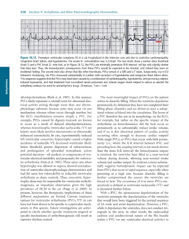
              Figure 18.13.  Premature	ventricular	complexes	(V)	in	a	cat	hospitalized	in	the	intensive	care	unit	due	to	restrictive	cardiomyopathy,
              congestive	heart	failure,	and	hypokalemia.	The	serum	K+	concentration	was	3.2	mEq/l.	The	two	insets	show	a	normal	sinus	heartbeat
              (inset	1)	and	a	PVC	(inset	2);	note	that,	as	in	Figure	18.12,	the	PVCs	are	minimally	premature	(R-R	interval:	red	bar	only	slightly	shorter
              than	blue	bar).	Thus,	the	hemodynamic	consequences	from	these	PVCs	would	be	expected	to	be	minimal,	and	indeed	they	were	an
              incidental	finding.	The	second	inset	also	shows	that	like	other	heartbeats,	PVCs	consist	of	a	QRS	and	a	T	wave.	Subjectively,	via	in-ICU
              telemetric	monitoring,	the	PVCs	decreased	substantially	in	number	with	correction	of	hypokalemia	and	congestive	heart	failure	alone.
              This	sequence	suggests	that	the	PVCs	may	have	been	caused	by	a	combination	of	cardiomyopathy,	hypokalemia,	and	pulmonary	edema-
      Arrhythmias  arrhythmia	without	the	need	for	antiarrhythmic	drugs.	25	mm/sec,	1	cm	=	1	mV.
              induced	hypoxemia,	and	that	treatment	that	normalized	serum	potassium	and	arterial	oxygen	levels	helped	to	reduce	or	abolish	the


              afterdepolarizations (Rials et al. 1995). In this manner,   The most meaningful impact of PVCs on the patient
              PVCs likely represent a catchall term for abnormal elec-  relates to diastolic filling. When the ventricles depolarize
              trical  activity  arising  through  more  than  one  electro-  prematurely, by definition they have not completed their
              physiologic substrate, because some may occur via one   filling phase (diastole) and are driven to eject a subop-
              mechanism whereas others occur through another, but   timal volume of blood into the circulation. The harm of
              the  ECG  manifestation  remains  simply  a  PVC.  For   a PVC therefore lies not in its morphology on the ECG,
              example, PVCs caused by digoxin toxicosis are known   for  example,  but  rather  on  the  specific  impact  of  the
              to  occur  as  a  result  of  delayed  afterdepolarizations,   arrhythmia  on  hemodynamics:  did  the  beat  occur  so
              whereas monomorphic ventricular tachycardia (VT; see   prematurely  as  to  substantially  reduce  stroke  volume,
              below) more likely involves microreentry or abnormally   and  if  so,  is  this  abnormal  pattern  of  cardiac  activity
              enhanced automaticity. In cats, experimentally induced   occurring  often  enough  to  decrease  cardiac  output?
              left ventricular concentric hypertrophy caused a higher   With single PVCs, or PVCs that occur with little prema-
              incidence of inducible VT, decreased ventricular fibril-  turity  (i.e.,  where  the  R-R  interval  between  PVC  and
              lation  threshold,  greater  dispersion  of  refractoriness,   preceding beat, the coupling interval, is not much shorter
              and  prolongation  of  epicardial  monophasic  action   than the sinus R-R interval) the hemodynamic impact
              potential duration—all markers or components of ven-  is minimal: the ventricles have filled to a near-normal
              tricular electrical instability and propensity for ventricu-  volume  during  diastole,  allowing  near-normal  stroke
              lar arrhythmia (Rials et al. 1995). These same cats, when   volume and cardiac output. By contrast, a more substan-
              hypertrophy was allowed to regress, showed normaliza-  tially  negative  hemodynamic  impact  can  be  expected
              tion of ventricular electrophysiologic characteristics and   from PVCs that occur in rapid sequence, especially when
              had the same low vulnerability to inducible ventricular   persisting  at  a  high  rate,  because  diastolic  filling  is
              arrhythmia as sham controls. Thus, concentric hyper-  further  compromised  the  sooner  the  ventricles  are
              trophy alone may be responsible for ventricular arrhyth-  forced to beat. The occurrence of 4 or more PVCs con-
              mogenesis,  an  important  observation  given  the  high   secutively is defined as ventricular tachycardia (VT) and
              prevalence  of  HCM  in  the  cat  (Paige  et  al.  2009).  At   is discussed further below.
              present, however, the therapeutic implications of these   With a PVC, the spontaneous depolarization of the
              different  mechanisms  are  limited.  Current  treatment   ventricles preempts the depolarization of the ventricles
              options for ventricular arrhythmias (PVCs, VT) in cats   that would have been triggered by the normal sequence
              have not been shown to be specific to a particular mech-  of SA node and atrial depolarization. However, a PVC,
              anism  in  this species. Future  investigations are  antici-  while it depolarizes the ventricles, does not conduct ret-
              pated to clarify whether specific treatments targeted at   rograde  to  the  atria.  In  other  words,  the  endocardial
              specific mechanisms of arrhythmogenesis will result in   cushion  and  unidirectional  nature  of  the  His  bundle
              superior rhythm control.                           isolate a PVC (or any ventricular electrical activity) to

