Page 226 - Feline Cardiology
P. 226
Chapter 18: Arrhythmias and Other Electrocardiographic Abnormalities  231


              according to site of origin: a PVC that begins in the left   inaccurate prognosis, or otherwise bring harm. The dif-
              ventricular  free  wall  spreads  toward  the  right,  a  very   ferential diagnosis for wide, bizarre QRS complexes on
              different pattern from normal, resulting in a very wide   an ECG consists of
              and  different-appearing  QRS  complex.  By  contrast,  a
              PVC that originates near the point of entry of the His   •  Premature ventricular complex/ventricular tachycardia/
              bundle into the ventricles (e.g., base of the right ven-  AV dissociation with accelerated junctional rhythm
              tricle) may spread through the ventricles in a manner   •  Ventricular escape rhythm (Figure 18.20)
              not substantially different from normal depolarization,   •  Sinus rhythm with right or left bundle branch block
              and the morphology of such a PVC is only mildly dif-  or fascicular block (Figure 18.22)
              ferent from the normal QRS complex (see Figure 18.12).  •  Right axis deviation due to right ventricular enlarge-
                 Several  electrocardiographic  impostors  exist  that   ment or displacement of the heart (Figure 18.16)
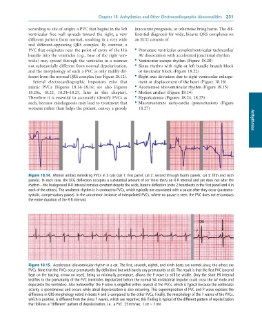
              mimic  PVCs  (Figures  18.14–18.16;  see  also  Figures   •  Accelerated idioventricular rhythm (Figure 18.15)
              18.20a,  18.22,  18.24–18.27,  later  in  this  chapter).   •  Motion artifact (Figure 18.14)
              Therefore it is essential to accurately identify PVCs as   •  Hyperkalemia (Figures 18.24, 18.25)
              such, because misdiagnosis may lead to treatment that   •  Macroreentrant  tachycardia  (preexcitation)  (Figure
              worsens rather than helps the patient, convey a grossly   18.27)                                          Arrhythmias


               Figure 18.14.  Motion	artifact	mimicking	PVCs	in	3	cats	(cat	1:	first	panel;	cat	2:	second	through	fourth	panels;	cat	3:	fifth	and	sixth
               panels).	In	each	case,	the	ECG	deflection	occupies	a	substantial	amount	of	(or	more	than)	an	R-R	interval	and	yet	does	not	alter	the
               rhythm—the	background	R-R	interval	remains	constant	despite	the	wide,	bizarre	deflection	(note	2	heartbeats	in	the	first	panel	and	4	in
               each	of	the	others).	The	unaltered	rhythm	is	in	contrast	to	PVCs,	which	typically	are	associated	with	a	pause	after	they	occur	(postextra-
               systolic,	compensatory	pause).	In	the	uncommon	instance	of	interpolated	PVCs,	where	no	pause	is	seen,	the	PVC	does	not	encompass
               the	entire	duration	of	the	R-R	interval.


                                                                                               QRS

                                                                                              P
                                                                                                    T


               Figure 18.15.  Accelerated	idioventricular	rhythm	in	a	cat.	The	first,	seventh,	eighth,	and	ninth	beats	are	normal	sinus;	the	others	are
               PVCs.	Note	that	the	PVCs	occur	prematurely	(by	definition)	but	with	barely	any	prematurity	at	all.	The	result	is	that	the	first	PVC	(second
               beat	on	the	tracing;	arrow	on	inset),	being	so	minimally	premature,	allows	the	P	wave	to	still	be	visible.	Only	the	short	PR	interval
               testifies	to	the	prematurity	of	the	PVC	(ventricles	depolarized	before	the	normal	SA	nodal/atrial	impulse	could	cross	the	AV	node	and
               depolarize	the	ventricles).	Also	noteworthy:	the	P	wave	is	engulfed	within	several	of	the	PVCs,	which	is	typical	because	the	ventricular
               activity	is	spontaneous	and	occurs	while	atrial	depolarization	is	also	occurring.	This	superimposition	of	PVC	and	P	wave	explains	the
               difference	in	QRS	morphology	noted	in	beats	4	and	5	compared	to	the	other	PVCs.	Finally,	the	morphology	of	the	T	waves	of	the	PVCs,
               which	is	positive,	is	different	from	the	sinus	T	waves,	which	are	negative;	this	finding	is	typical	of	the	different	pattern	of	repolarization
               that	follows	a	“different”	pattern	of	depolarization,	i.e.,	a	PVC.	25	mm/sec,	1	cm	=	1	mV.

