Page 229 - Feline Cardiology
P. 229
234  Section F: Arrhythmias and Other Electrocardiographic Abnormalities


                                                                                                     *

                                                                                           QRS
                                                                                          P

                                                                                               T


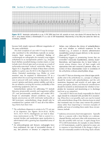
              Figure 18.17.  Ventricular	tachycardia	in	a	cat.	A	PVC	(fifth	beat	from	left;	asterisk	on	inset;	note	shorter	R-R	interval	than	for	the
              first	4,	sinus	beats)	initiates	a	monomorphic	VT	at	a	rate	of	300	beats/minute.	Macroreentry	is	less	likely	but	cannot	be	ruled	out.
              25	mm/sec,	5	mm/mV.


              because both simply represent different magnitudes of   failure, may influence the choice of antiarrhythmics
      Arrhythmias  cific (unrelated to the arrhythmia; typically an asymp-  moment  (e.g.,  if  centesis  or  diuretic  administration
                                                                   and  even  whether  to  withhold  treatment  for  the
              the same arrhythmia.
                 The chief complaint of cats with VT may be nonspe-
                                                                   reestablishes normal oxygen delivery to the myocar-
                                                                   dium and VT resolves).
              tomatic  heart  murmur  or  incidental  finding  of
              cardiomegaly on radiographs, for example), may suggest  •  Underlying  triggers  or  potentiating  factors  that  are
              arrhythmia in an asymptomatic patient (e.g., irregular   reversible—classically hypokalemia, anemia, hyper-
              heart rhythm ausculted during a routine exam), or may   thyroidism,  and  hypoxemia  due  to  heart  failure  or
              include overt clinical signs such as syncope as a result of   severe  airway  disease—have  all  been  sought  with
              tachycardia-associated  reduced  ventricular  filling  (see   appropriate tests and corrected if present; often, cor-
              Figure 18.1). Regardless of which of these three contexts   rection of these abnormalities leads to resolution of
              applies to a given case, the ECG is the diagnostic test of   the arrhythmia without antiarrhythmic drugs.
              choice.  Extended  monitoring  (e.g.,  Holter  or  event
              monitor)  may  be  required  to  demonstrate  VT  in  a   Cats with VT that are showing overt clinical signs attrib-
              patient where it is occurring intermittently, and the deci-  utable only to the arrhythmia (i.e., syncope or collapse)
              sion to proceed with such testing is dependent on the   should  be  treated  with  antiarrhythmics,  and  the  cat’s
              owner’s opinion and desire, patient stability and risk to   mentation,  frequency  and  severity  of  such  signs,  and
              the cat related to carrying the monitor, availability of the   owner perception, desires, and financial considerations
              monitoring equipment, and cost.                    should be included as determinants for whether to hos-
                 Antiarrhythmic  options  for  addressing  VT  include   pitalize  for  treatment  and  monitoring  or  to  discharge
              lidocaine, propranolol, esmolol, and magnesium sulfate   and treat as an outpatient.
              (IV)  and  procainamide,  mexiletine,  and  sotalol  (PO).   Cats with VT that is not clearly responsible for clinical
              Except  in  catastrophic  clinical  conditions  (e.g.,  col-  signs should be evaluated with portable ECG monitor-
              lapsed, unconscious cat with ventricular tachycardia at   ing. When this is not possible, such cats may be treated
              340  beats/min),  antiarrhythmic  drugs  should  only  be   with antiarrhythmic drugs if the clinician keeps in mind
              considered for a patient with VT once all of the follow-  that treatment may in some cases worsen the patient’s
              ing criteria have been met:                        status rather than improving it. Common examples of
                                                                 pitfalls, and ways of avoiding them under these circum-
              •  The arrhythmia diagnosis is certain to be VT, not any   stances, include
                of  its  impostors  (see  Figures  18.14–18.16,  18.20a,
                18.22,  18.24–18.27);  misdiagnosis  can  lead  to  disas-  •  Judicious, not excessive, heart rate suppression.  Typically
                trous mistreatment as the “VT” fails to convert to a   this  is  a  concern  with  a  beta  blocker  or  a  calcium-
                normal rhythm and medication upon medication is    channel blocker, and most often with intravenous use
                administered with a growing but unjustified sense of   but also possibly with oral use. A rapid change from
                urgency.                                           VT at 260 beats/minute, for example, to sinus rhythm
              •  An  echocardiogram  and  thoracic  radiographs  have   at 150 beats/minute, could be expected in some cats
                been  performed;  the  type  of  myocardial  disease,  if   in response to beta-blockade if the dosage is high or
                any,  and  presence  or  absence  of  congestive  heart   the cat is unusually sensitive to the drug. Such a drastic

