Page 233 - Feline Cardiology
P. 233
238  Section F: Arrhythmias and Other Electrocardiographic Abnormalities


              in the AV node and is said to carry a good prognosis   undertaken  using  an  atropine  response  test:  an  ECG
              because it is closely related to first-degree AV block and   strip  is  recorded,  and  0.04 mg/kg  atropine  is  immedi-
              virtually never causes clinical signs. This form of second-  ately  administered  IV.  One  or  both  follow-up  ECGs
              degree AV block is very uncommon in the cat, perhaps   recorded  15  and  30  minutes  later  should  show  sinus
              because, like first-degree AV block, it usually arises in   tachycardia  when  second-degree  AV  block  is  vagally
              states of parasympathetic predominance. The other type   mediated  and  therefore  likely  harmless;  this  is  more
              of second-degree AV block, Mobitz type II, by contrast   likely with Mobitz type I than II, but either type may
              demonstrates perfectly regular PR intervals for all QRS   respond to atropine. If atropine response is noted, and
              complexes, until suddenly one or more P wave(s) is/are   the cat displayed symptoms consistent with poor arterial
              blocked. Mobitz type II second-degree AV block arises   perfusion, such as syncope, treatment with terbutaline
              from the AV bundle and is said to carry a more guarded   may  be  tried  (0.625 mg  PO  q  8–12h);  propantheline
              to  poor  prognosis  because  it  more  closely  resembles   (0.8–1.6 mg/kg PO q 8h, titrate down or stop if constipa-
              third-degree AV  block.  However,  objective  evidence  is   tion  or  other  extracardiac  vagolytic  signs  occur)  is  a
              lacking  to  support  this  extrapolation  of  severity  in   second choice if terbutaline is not effective or not toler-
              Mobitz types I and II from human cardiology to feline   ated. However, many syncopal cats with AV block ulti-
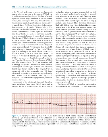
              patients. In “simple” Mobitz type II second-degree AV   mately  may  require  a  pacemaker  (see  below).  In  the
      Arrhythmias  waves,  whereas  in “advanced”  or “high  grade”  Mobitz   syncope, treatment of second-degree AV block generally
                                                                 absence  of  overt  clinical  signs  such  as  weakness  or
              block, more conducted P waves occur than blocked P
                                                                 is  not  necessary.  An  exception  to  this  rule  is  a  stable
              type  II  second-degree  AV  block  (Figure  18.19),  more
              blocked  P  waves  occur  than  conducted  P  waves.  The
              presence or absence of clinical signs in second-degree   patient that requires general anesthesia, where bradycar-
                                                                 dia that is unresponsive to anticholinergics may develop
              AV block appears to be related to the overall ventricular   intraoperatively. Cats with high-grade second-degree AV
              rate. Therefore Mobitz type I second-degree AV block   block should be instrumented with a temporary pace-
              essentially never produces clinical manifestations such   maker (Côté and Laste 2000; Petrie 2005) if they require
              as  lethargy  or  syncope,  whereas  the  more  advanced   general  anesthesia,  as  they  may  otherwise  arrest  or
              Mobitz type II second-degree AV block cases often have   develop severe bradycardia at induction or during main-
              clinical signs that are similar to third-degree AV block,   tenance, and drugs that only accelerate SA nodal activity
              concurrent  with  a  slow  ventricular  rate  (100  beats/  (atropine, glycopyrrolate, epinephrine) are likely to be
              minute or less): weakness, lethargy, syncope, and cardio-  ineffective  because  they  rarely  increase  conduction
              genic  seizures  (true  convulsions  caused  by  critical,   speed through a diseased AV node. In second-degree AV
              bradycardia-induced cerebral hypoperfusion) even with   block,  drugs  with  negative  chronotropic  properties,
              minimal exertion.                                  including beta blockers and calcium-channel blockers,
                 Assessment  of  the  physiologic  (generally  harmless)   are contraindicated.
              versus  pathologic  (may  cause  clinical  signs  such  as   Third-degree  AV  block  is  a  complete  and  sustained
              syncope)  nature  of  second-degree  AV  block  may  be   interruption  of  AV  conduction.  The  only  ventricular


                  *       *       *


              Figure 18.19.  Mobitz	type	II	second-degree	AV	block,	3	:	1,	in	a	cat.	The	first	beat	on	the	left	is	a	sinus	beat,	with	P	wave	(1st	asterisk)
              followed	by	a	QRS.	The	second	P	wave	is	blocked	(2nd	asterisk)	and	no	ventricular	activity	is	present.	The	third	P	wave	(3rd	asterisk)
              is	preempted	by	a	ventricular	escape	beat,	which	emerges	because	the	ventricles	have	been	inactive	for	0.44	sec	(the	time	from	the
              normal	sinus	beat	to	the	escape	beat),	an	effective	ventricular	escape	rate	of	136	beats/minute.	This	pattern—1	normal	sinus	beat,	then
              2	blocked	P	waves	with	a	ventricular	escape	beat	occurring	around	the	time	of	the	second	blocked	P	wave—repeats	for	the	rest	of	the
              tracing.	50	mm/sec,	1	cm	=	1	mV.

