Page 236 - Feline Cardiology
P. 236
Chapter 18: Arrhythmias and Other Electrocardiographic Abnormalities  241


                     A                                               B                                                  Arrhythmias


                                             P
                       P      P                            P      P              P      P      P      P       P
                  QRS
                                                                                          QRS    QRS    QRS
                         QRS

              C
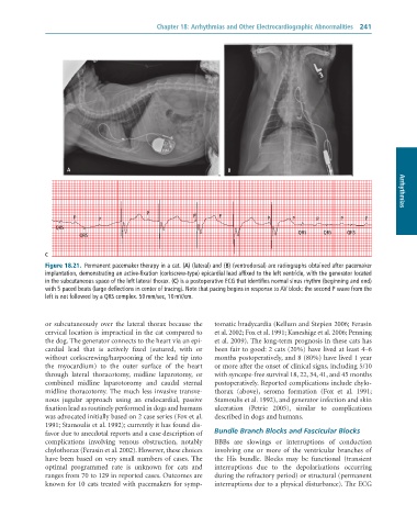
              Figure 18.21.  Permanent	pacemaker	therapy	in	a	cat.	(A)	(lateral)	and	(B)	(ventrodorsal)	are	radiographs	obtained	after	pacemaker
              implantation,	demonstrating	an	active-fixation	(corkscrew-type)	epicardial	lead	affixed	to	the	left	ventricle,	with	the	generator	located
              in	the	subcutaneous	space	of	the	left	lateral	thorax.	(C)	is	a	postoperative	ECG	that	identifies	normal	sinus	rhythm	(beginning	and	end)
              with	5	paced	beats	(large	deflections	in	center	of	tracing).	Note	that	pacing	begins	in	response	to	AV	block:	the	second	P	wave	from	the
              left	is	not	followed	by	a	QRS	complex.	50	mm/sec,	10	mV/cm.


              or  subcutaneously  over  the  lateral  thorax  because  the   tomatic bradycardia (Kellum and Stepien 2006; Ferasin
              cervical location is impractical in the cat compared to   et al. 2002; Fox et al. 1991; Kaneshige et al. 2006; Penning
              the dog. The generator connects to the heart via an epi-  et al. 2009). The long-term prognosis in these cats has
              cardial  lead  that  is  actively  fixed  (sutured,  with  or   been fair to good: 2 cats (20%) have lived at least 4–6
              without  corkscrewing/harpooning  of  the  lead  tip  into   months postoperatively, and 8 (80%) have lived 1 year
              the  myocardium)  to  the  outer  surface  of  the  heart   or more after the onset of clinical signs, including 5/10
              through  lateral  thoracotomy,  midline  laparotomy,  or   with syncope-free survival 18, 22, 34, 41, and 45 months
              combined  midline  laparotoromy  and  caudal  sternal   postoperatively. Reported complications include chylo-
              midline thoracotomy. The much less invasive transve-  thorax  (above),  seroma  formation  (Fox  et  al.  1991;
              nous  jugular  approach  using  an  endocardial,  passive   Stamoulis et al. 1992), and generator infection and skin
              fixation lead as routinely performed in dogs and humans   ulceration  (Petrie  2005),  similar  to  complications
              was advocated initially based on 2 case series (Fox et al.   described in dogs and humans.
              1991; Stamoulis et al. 1992); currently it has found dis-
              favor due to anecdotal reports and a case description of   Bundle Branch Blocks and Fascicular Blocks
              complications  involving  venous  obstruction,  notably   BBBs  are  slowings  or  interruptions  of  conduction
              chylothorax (Ferasin et al. 2002). However, these choices   involving  one  or  more  of  the  ventricular  branches  of
              have been based on very small numbers of cases. The   the  His  bundle.  Blocks  may  be  functional  (transient
              optimal  programmed  rate  is  unknown  for  cats  and   interruptions  due  to  the  depolarizations  occurring
              ranges from 70 to 129 in reported cases. Outcomes are   during the refractory period) or structural (permanent
              known for 10 cats treated with pacemakers for symp-  interruptions due to a physical disturbance). The ECG

