Page 232 - Feline Cardiology
P. 232
Chapter 18: Arrhythmias and Other Electrocardiographic Abnormalities  237


              a period of at least 15 minutes if no benefit is observed,   form of excessive restriction of conduction to the ven-
              and  then  followed  with  esmolol  or  propranolol  if  2     tricles, called AV block. AV blocks are defined as delays
              doses of lidocaine were ineffective. Refractory VT is the   or stoppages of conduction between the atria and the
              diagnosis  of  exclusion  if  no  improvement  is  observed   ventricles.
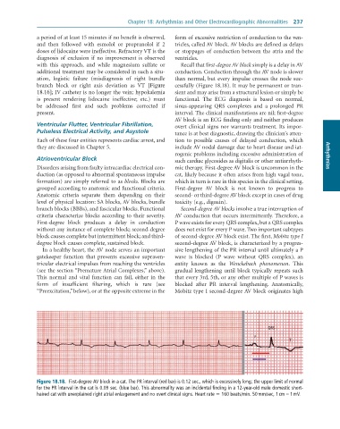
              with  this  approach,  and  while  magnesium  sulfate  or   Recall that first-degree AV block simply is a delay in AV
              additional treatment may be considered in such a situ-  conduction. Conduction through the AV node is slower
              ation,  logistic  failure  (misdiagnosis  of  right  bundle   than normal, but every impulse crosses the node suc-
              branch  block  or  right  axis  deviation  as  VT  [Figure   cessfully (Figure 18.18). It may be permanent or tran-
              18.16]; IV catheter is no longer the vein; hypokalemia   sient and may arise from a structural lesion or simply be
              is  present  rendering  lidocaine  ineffective;  etc.)  must    functional.  The  ECG  diagnosis  is  based  on  normal,
              be  addressed  first  and  such  problems  corrected  if     sinus-appearing  QRS  complexes  and  a  prolonged  PR
              present.                                           interval. The clinical manifestations are nil; first-degree
                                                                 AV block is an ECG finding only and neither produces
              Ventricular Flutter, Ventricular Fibrillation,     overt clinical signs nor warrants treatment. Its impor-
              Pulseless Electrical Activity, and Asystole        tance is at best diagnostic, drawing the clinician’s atten-
              Each of these four entities represents cardiac arrest, and   tion  to  possible  causes  of  delayed  conduction,  which
              they are discussed in Chapter 5.                   include AV nodal damage due to heart disease and iat-
                                                                 rogenic problems including excessive administration of
              Atrioventricular Block                             such cardiac glycosides as digitalis or other antiarrhyth-  Arrhythmias
              Disorders arising from faulty intracardiac electrical con-  mic therapy. First-degree AV block is uncommon in the
              duction (as opposed to abnormal spontaneous impulse   cat, likely because it often arises from high vagal tone,
              formation) are simply referred to as blocks. Blocks are   which in turn is rare in this species in the clinical setting.
              grouped according to anatomic and functional criteria.   First-degree  AV  block  is  not  known  to  progress  to
              Anatomic  criteria  separate  them  depending  on  their   second- or third-degree AV block except in cases of drug
              level of physical location: SA blocks, AV blocks, bundle   toxicity (e.g., digoxin).
              branch blocks (BBBs), and fascicular blocks. Functional   Second-degree AV blocks involve a true interruption of
              criteria characterize blocks according to their severity.   AV conduction that occurs intermittently. Therefore, a
              First-degree  block  produces  a  delay  in  conduction   P wave exists for every QRS complex, but a QRS complex
              without any instance of complete block; second-degree   does not exist for every P wave. Two important subtypes
              block causes complete but intermittent block; and third-  of second-degree AV block exist. The first, Mobitz type I
              degree block causes complete, sustained block.     second-degree AV block, is characterized by a progres-
                 In a healthy heart, the AV node serves an important   sive lengthening of the PR interval until ultimately a P
              gatekeeper  function  that  prevents  excessive  supraven-  wave  is  blocked  (P  wave  without  QRS  complex),  an
              tricular electrical impulses from reaching the ventricles   entity  known  as  the  Wenckebach  phenomenon.  This
              (see the section “Premature Atrial Complexes,” above).   gradual  lengthening  until  block  typically  repeats  such
              This  normal  and  vital  function  can  fail,  either  in  the   that every 3rd, 5th, or any other multiple of P waves is
              form  of  insufficient  filtering,  which  is  rare  (see   blocked  after  PR  interval  lengthening.  Anatomically,
              “Preexcitation,” below), or at the opposite extreme in the   Mobitz type I second-degree AV block originates high


                                                                                                   QRS
                                                                                              P
                                                                                                           T


              Figure 18.18.  First-degree	AV	block	in	a	cat.	The	PR	interval	(red	bar)	is	0.12	sec.,	which	is	excessively	long;	the	upper	limit	of	normal
              for	the	PR	interval	in	the	cat	is	0.09	sec.	(blue	bar).	This	abnormality	was	an	incidental	finding	in	a	12-year-old	male	domestic	short-
              haired	cat	with	unexplained	right	atrial	enlargement	and	no	overt	clinical	signs.	Heart	rate	=	160	beats/min.	50	mm/sec,	1	cm	=	1	mV.

