Page 234 - Feline Cardiology
P. 234
Chapter 18: Arrhythmias and Other Electrocardiographic Abnormalities  239


                                                                                                           P
                                                                                      P      P
                                                                                                    P      +
                                                                                                           T
                                                                                        T
                                                                                                  T


                                                                                    QRS
                                                                                             QRS
                                                                                                       QRS

               A


                                                         P   P    P   P                                                 Arrhythmias


               B
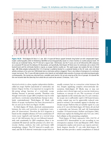
              Figure 18.20.  Third-degree	AV	block	in	2	cats.	(A)	A	16-year-old	female	spayed	domestic	long-haired	cat	with	compensated	hyper-
              trophic	cardiomyopathy	(mild	LV	thickening)	identified	echocardiographically	based	on	a	heart	murmur	on	routine	physical	exam.	AV
              block	was	an	incidental	finding.	The	P-P	interval	is	regular	(rate:	230/minute),	but	the	P	waves	are	not	all	followed	by	QRS	complexes
              at	a	consistent	PR	interval;	this	indicates	AV	dissociation,	and	specifically,	third-degree	AV	block	since	the	ventricular	rate	is	slower	(150
              beats/minute)	and	the	ventricular	rhythm	is	regular,	as	escape	rhythms	usually	are.	This	rapid	escape	rate	explains	the	lack	of	clinical
              signs.	(B)	Wide-complex	tachycardia	(first	1/3	of	tracing)	is	suddenly	blocked	in	a	sustained	fashion	at	the	AV	node,	such	that	atrial
              activity	continues	at	the	same	rate	(180/minute)	but	no	ventricular	activity	is	triggered.	There	is	also	failure	of	emergence	of	a	ventricular
              escape	mechanism.	This	13-year-old	male	domestic	short-haired	cat	had	multiple	daily	episodes	of	syncope	and	underlying	hypertrophic
              cardiomyopathy.	This	tracing	was	obtained	from	a	portable	event	monitor	the	cat	was	wearing	at	the	time	of	syncope.	An	episode	like
              this	one	occurred	later	the	same	day	from	which	the	cat	could	not	be	revived.	25	mm/sec,	1	cm	=	1	mV.


              electrical activity is a slow, regular, independent rhythm,   usually constant, but no connection exists between the
              called an escape rhythm (junctional or ventricular; see   two.  Despite  signifying  complete  atrioventricular  dis-
              below) (Figure 18.20a). It is important to recognize the   sociation,  third-degree  AV  blocks  may  or  may  not
              life-saving  salvage  function  of  a  ventricular  escape   produce overt clinical signs such as exercise intolerance,
              rhythm,  because  it  prevents  asystole.  Therefore  even   weakness,  or  syncope  in  cats.  The  occurrence  of  such
              though ventricular escape QRS complexes may be wide   signs depends on the ventricular rate. If a cat’s ventricu-
              and bizarre, ventricular antiarrhythmic therapy is abso-  lar escape rate is sufficiently rapid (>100 beats/minute),
              lutely  contraindicated.  Third-degree  AV  block  with   cardiac  output  may  not  be  appreciably  reduced  com-
              failure of escape mechanisms has been documented in   pared to normal. Cats routinely appear to develop ven-
              the cat and can be fatal (see Figure 18.20b).      tricular escape rhythms that are suitably rapid: in a case
                 In third-degree AV block, electrical communication   series of 21 cats with third-degree AV block, all cats had
              between  the  atria  and  ventricles  is  nonexistent  (com-  a  ventricular  rate  between  100  and  140  beats/minute
              plete AV dissociation). Therefore the ECG diagnosis is   (median 120 beats/minute) (Kellum and Stepien 2006),
              based on the complete absence of P wave conduction (P   which may explain the finding that only 3/21 cats (14%)
              waves  occur  regularly  and  typically  at  a  normal  sinus   had collapse as the presenting complaint.
              rate of 140–260/minute, but are not followed immedi-  Third-degree AV block represents 5.8% (10/170) of
              ately  and  consistently  by  QRS  complexes;  there  is  no   recorded  ECG  abnormalities  in  cats  according  to  one
              consistent PR interval) and the presence of a slow ven-  retrospective analysis (Côté and Harpster 2009). Three
              tricular  rhythm,  uniform,  but  wide  and  bizarre  QRS   retrospective studies and several case reports and text-
              morphology  (shape).  The  P-P  interval  is  usually  con-  book  chapters  describe  37  cats  with  third-degree  AV
              stant, and the R-R interval (ventricular escape beats) is   block and thus provide a useful compilation of clinical

