Page 238 - Feline Cardiology
P. 238
Chapter 18: Arrhythmias and Other Electrocardiographic Abnormalities  243


              finding, especially if the diagnosis was reached partly or   atrial myopathy and hyperkalemia using the ECG alone
              entirely in error due to incorrect limb position during   is difficult. If P waves are not evident, immediate mea-
              restraint for the ECG (see Chapter 9). Clinical manifes-  surement of serum electrolytes is warranted.
              tations of BBBs alone generally do not occur. These dis-
              turbances  therefore  do  not  warrant  specific  treatment   Sinus Node Dysfunction (Sick Sinus Syndrome,
              beyond that of their underlying cause if one exists. The   Bradycardia-Tachycardia Syndrome)
              importance of recognizing BBB lies in the fact that they   This polyarrhythmic disorder, which is well-recognized
              could  be  the  first  indicators  of  underlying  cardiac   in humans and dogs, has never been reported as such in
              disease,  which  itself  warrants  further  diagnosis  and   the  cat.  An  autoimmune  disturbance  directed  at  the
              treatment, and that they can—and should not—be mis-  heart’s nodal tissues, or at autonomic receptors, has been
              interpreted as ventricular arrhythmias.            shown in humans with this disorder, but such mecha-
                                                                 nisms have not been evaluated in veterinary medicine
              Atrial Standstill (Silent Atrium)                  (Côté 2010). The hallmark ECG findings are a mixture
              This  rhythm  disturbance  is  characterized  by  the  total   of  sinus  pause/sinus  arrest  with  a  failure  of  escape
              absence of atrial depolarization. The three differential   rhythm  to  emerge  (causing  asystole  of  up  to  several
              diagnoses for atrial standstill are 1) moderate to marked   seconds’ duration) and AV block, as bradycardias; PVCs
                             +
              hyperkalemia  (K   >7.5 mEq/l;  see  below),  2)  atrial   and/or PACs as tachycardias; and generally a variety of
              myopathy  (Gavaghan  et  al.  1999;  Richig  and  Tilley   these  arrhythmias  occurring  unpredictably  and  some-  Arrhythmias
              1984), and 3) ECG artifact (P waves too small, or iso-  times simultaneously on the same ECG strip. Based on
              electric, preventing them from being seen properly on a   cardiac event monitoring in dogs, it is bradycardia that
              chosen ECG lead). Given the very small size of normal   is associated with the clinical signs, and therefore optimal
              feline P waves, a 10-lead ECG (with reduced or no noise   treatment includes pacemaker implantation. The prog-
              filtration and an increased gain of 20 mm/mv [Schrope   nosis in cats is unknown, as it has not been reported in
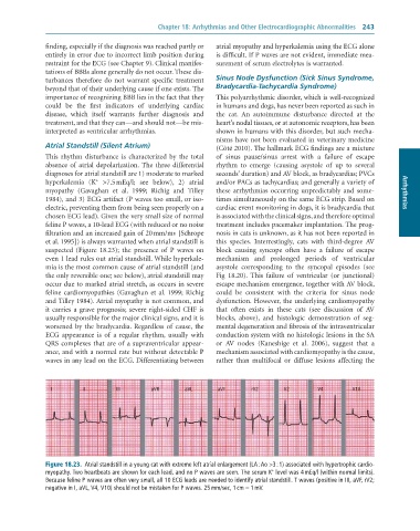
              et al. 1995]) is always warranted when atrial standstill is   this  species.  Interrestingly,  cats  with  third-degree  AV
              suspected  (Figure  18.23);  the  presence  of  P  waves  on   block  causing  syncope  often  have  a  failure  of  escape
              even 1 lead rules out atrial standstill. While hyperkale-  mechanism  and  prolonged  periods  of  ventricular
              mia is the most common cause of atrial standstill (and   asystole  corresponding  to  the  syncopal  episodes  (see
              the only reversible one; see below), atrial standstill may   Fig  18.20).  This  failure  of  ventricular  (or  junctional)
              occur due to marked atrial stretch, as occurs in severe   escape mechanism emergence, together with AV block,
              feline  cardiomyopathies  (Gavaghan et  al.  1999; Richig   could  be  consistent  with  the  criteria  for  sinus  node
              and Tilley 1984). Atrial myopathy is not common, and   dysfunction. However, the underlying cardiomyopathy
              it carries a grave prognosis; severe right-sided CHF is   that  often  exists  in  these  cats  (see  discussion  of  AV
              usually responsible for the major clinical signs, and it is   blocks,  above),  and  histologic  demonstration  of  seg-
              worsened  by  the  bradycardia.  Regardless  of  cause,  the   mental degeneration and fibrosis of the intraventricular
              ECG  appearance  is  of  a  regular  rhythm,  usually  with   conduction system with no histologic lesions in the SA
              QRS complexes that are of a supraventricular appear-  or  AV  nodes  (Kaneshige  et  al.  2006),  suggest  that  a
              ance, and with a normal rate but without detectable P   mechanism associated with cardiomyopathy is the cause,
              waves in any lead on the ECG. Differentiating between   rather  than  multifocal  or  diffuse  lesions  affecting  the


                I        II        III        aVR       aVL      aVF       rV2       V2        V4        V10


              Figure 18.23.  Atrial	standstill	in	a	young	cat	with	extreme	left	atrial	enlargement	(LA	:	Ao	>3	:	1)	associated	with	hypertrophic	cardio-
                                                                                 +
              myopathy.	Two	heartbeats	are	shown	for	each	lead,	and	no	P	waves	are	seen.	The	serum	K 	level	was	4	mEq/l	(within	normal	limits).
              Because	feline	P	waves	are	often	very	small,	all	10	ECG	leads	are	needed	to	identify	atrial	standstill.	T	waves	(positive	in	III,	aVF,	rV2;
              negative	in	I,	aVL,	V4,	V10)	should	not	be	mistaken	for	P	waves.	25	mm/sec,	1	cm	=	1	mV.

