Page 243 - Feline Cardiology
P. 243
248  Section F: Arrhythmias and Other Electrocardiographic Abnormalities


                                                         T
                                                                                                           T


                                                                                                      QRS
                                                     QRS
                                                                        +
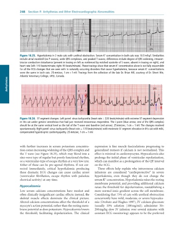
              Figure 18.25.  Hyperkalemia	in	2	male	cats	with	urethral	obstruction.	Serum	K 	concentration	in	both	cats	was	10.5	mEq/l.	Similarities
              include	atrial	standstill	(no	P	waves),	wide	QRS	complexes,	and	peaked	T	waves;	differences	include	degree	of	QRS	widening,	intraven-
              tricular	conduction	disturbance	(present	in	tracing	on	left	as	evidenced	by	notched	upstroke	of	S	wave,	absent	in	tracing	on	right),	and
                                                                                +
              heart	rate	(left:	140	beats/minute;	right:	90	beats/minute).	These	tracings	show	that	serum	K 	concentration	alone	is	not	fully	responsible
                                                                                                    +
              for	all	the	ECG	changes	that	are	seen	with	in	naturally	occurring	disorders	that	cause	hyperkalemia,	because	serum	K 	concentrations
              were	the	same	in	both	cats.	25	mm/sec,	1	cm	=	1	mV.	Tracings	from	the	collection	of	the	late	Dr.	Brian	Hill,	courtesy	of	Dr.	Sherri	Ihle,
              Atlantic	Veterinary	College,	UPEI,	Canada.
      Arrhythmias                                                                                       QRS


                                                      P
                                                          P
                                                                                                            T
                                                                                                       P


                                                       QRS

              Figure 18.26.  ST	segment	changes.	Left	panel:	sinus	tachycardia	(heart	rate	=	220	beats/minute)	with	extreme	ST	segment	depression
              in	this	cat	under	general	anesthesia	that	had	just	received	intravenous	meperidine.	The	J	point	(blue	arrow;	end	of	the	QRS	complex)
              should	be	at	the	same	vertical	level	as	the	tail	of	the	T	wave	and	baseline	(red	arrow).	25	mm/sec,	1	cm	=	1	mV.	The	changes	resolved
              spontaneously.	Right	panel:	sinus	tachycardia	(heart	rate	=	170	beats/minute)	with	moderate	ST	segment	elevation	in	this	cat	with	mild,
              compensated	hypertrophic	cardiomyopathy.	25	mm/sec,	1	cm	=	1	mV.


              with  further  increases  in  serum  potassium  concentra-  expression  is  fine  muscle  fasciculations  progressing  to
              tion comes increasing widening of the QRS complex and   generalized tremors if calcium is not normalized. This
              the T wave (see Figure 18.25), which may blend into a   effect is minimal in cardiomyocytes. Hypocalcemia also
              sine-wave type of regular but poorly functional rhythm,   prolongs the initial phase of ventricular repolarization,
              or a ventricular-type of escape rhythm at a very low rate.   which can manifest as a prolongation of the QT interval
              Either of these can be pre-agonal rhythms. If not cor-  on the ECG.
              rected  immediately,  critical  hyperkalemia  producing   These effects help explain why intravenous calcium
              these  dramatic  ECG  changes  can  cause  cardiac  arrest   infusions  are  considered  “cardioprotective”  in  severe
              (ventricular  fibrillation,  escape  rhythm  with  pulseless   hyperkalemia,  even  though  they  do  not  change  the
                                                                        +
              electrical activity) at any time.                  serum K  concentration. Hyperkalemia raises the resting
                                                                 membrane potential, and providing additional calcium
              Hypocalcemia                                       raises the threshold for depolarization, reestablishing a
              Low  serum  calcium  concentrations  have  modest  and   more normal ionic gradient across the cell membrane.
              often clinically insignificant cardiac effects; instead, the   Considering that 75% of cats with urethral obstruction
              skeletal  muscle  effects  dominate  the  clinical  picture.   concurrently have mild, moderate, or severe hypocalce-
              Altered calcium concentrations affect the threshold of a   mia (Drobatz and Hughes 1997), IV calcium gluconate
              myocyte’s action potential, rather than the resting mem-  (usually  10%  solution  [100 mg/ml];  administer  50–
              brane potential as does potassium. Hypocalcemia lowers   200 mg/kg slow IV infusion over several minutes with
              the  threshold,  facilitating  depolarization.  The  clinical   constant ECG monitoring) appears to be the preferred

